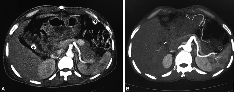
Fig. 4.

( A ) Computed tomography (CT) angiogram showing a pseudoaneurysm in relation to splenic artery in a patient post-Whipple's procedure. A safe coil deployment position could not be reached on digital subtraction angiography (DSA), so percutaneous thrombin injection performed. ( B ) Follow-up CT angiogram shows nonopacification of pseudoaneurysm.
