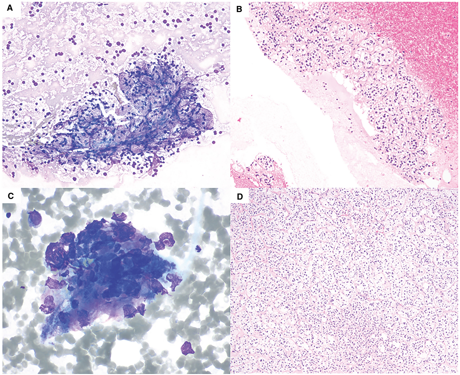
Figure 1.

Fine-needle aspiration of adrenal cortical adenoma. (A) Benign adrenal cortical elements with “frothy” background and bland-appearing naked nuclei. Intact cells have abundant clear foamy cytoplasms (Diff-Quik, 200×). (B) Cell block of this case shows nests of bland-appearing cells with abundant clear cytoplasm (H & E, 200×). (C) A case of atypia of undetermined significance (AUS) with crushed cells with occasional hyperchromatic nuclei (Diff-Quik, 600×). (D) The subsequent adrenalectomies of both cases showed adrenal cortical adenoma. The histological section shown is from the AUS case (H & E, 100×).
