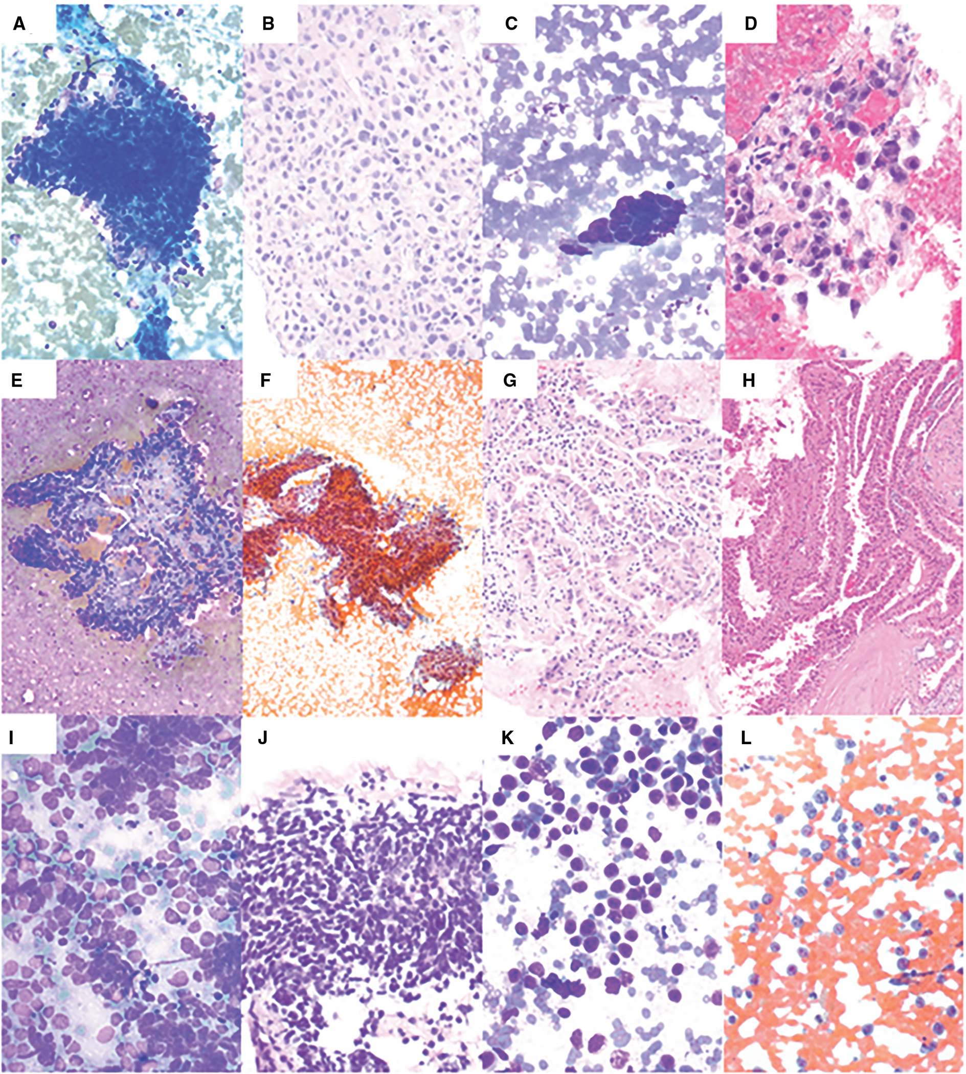
Figure 3.

Fine-needle aspiration of malignant neoplasms. (A) Adrenal cortical carcinoma showing nuclear overlapping, hyperchromasia, and nuclear pleomorphism (Diff-Quik, 200×). (B) Cell block shows pleomorphic cells with pale eosinophilic cytoplasm (H & E, 400×). (C) Poorly differentiated adenocarcinoma of the esophagus showing cohesive epithelial cells with nuclear pleomorphism (Diff-Quik, 400×). (D) Cell block shows pleomorphic epithelial cells forming a poorly defined gland and/or cluster (H & E, 400×). (E) Papillary renal cell carcinoma showing epithelial cells forming papillary structures with fibrovascular core (Diff-Quik, 100×). (F) Papanicolaou (PAP) stain shows epithelial cells forming papillary structures (PAP, 100×). (G) Cell block shows epithelial cells with abundant eosinophilic cytoplasm forming papillary structures (H & E, 200×). (H) The subsequent radical nephrectomy shows papillary renal cell carcinoma, type-2 (H & E, 100×). (I) Small cell carcinoma showing epithelioid cells with scant cytoplasm, fine chromatin, nuclear molding, and smearing artifact (Diff-Quik, 400×). (J) Cell block shows hyperchromatic epithelioid cells with crushing artifacts (H & E, 400×). (K) Diffuse large B-cell lymphoma showing discohesive, monotonous, large cells with scant cytoplasm (Diff-Quik, 400×). (L) PAP stain shows that these cells have occasional nucleoli (PAP, 400×).
