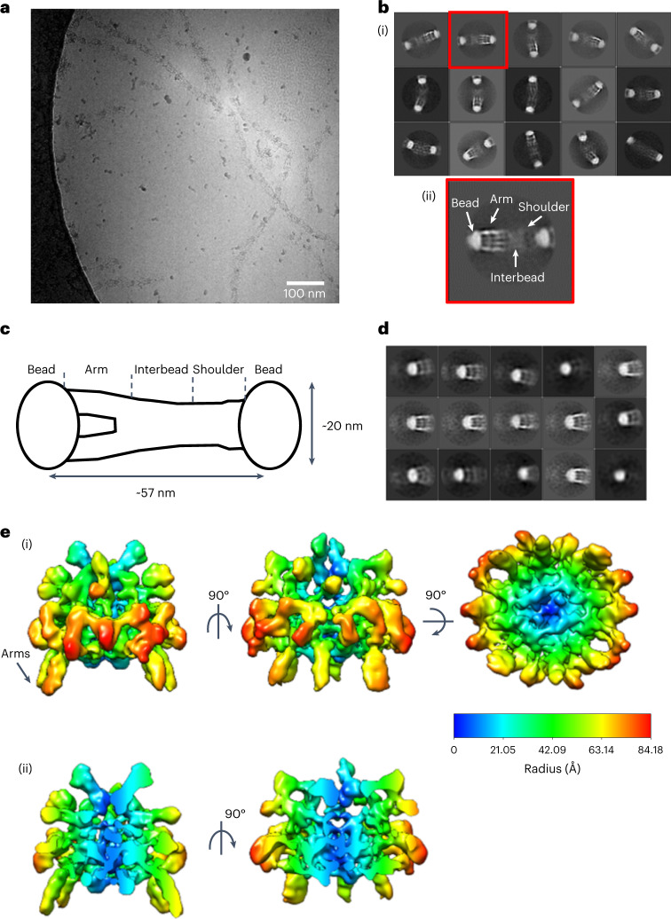
Fig. 1. Cryo-EM structure of the fibrillin microfibril bead region.
Fibrillin microfibrils were extracted from bovine ciliary zonules under nondenaturing conditions and imaged using cryo-EM. a, Representative cryo-EM image of purified bovine ciliary zonule microfibrils, from a dataset of 1,310 images. Scale bar, 100 nm. b, (i) Reference-free classification of the full fibrillin microfibril repeat. Box size, 100 nm. (ii) A class highlighted in panel (i) by a red box was rotated 180° and enlarged to highlight the different microfibril regions. c, Schematic of the fibrillin microfibril, highlighting the bead, arm, interbead and shoulder regions of the microfibril. d, Classification of particles aligned to the fibrillin bead region. Box size, 57 nm. e, (i) Cryo-EM single-particle reconstruction of the fibrillin bead region shown in three orthogonal orientations. The bead is rainbow colored by cylindrical radius from blue (at the center of the bead) to red (on the outside of the bead). (ii) Two orthogonal views of the bead reconstruction have been sliced to show a cross-section through the center of the bead.

