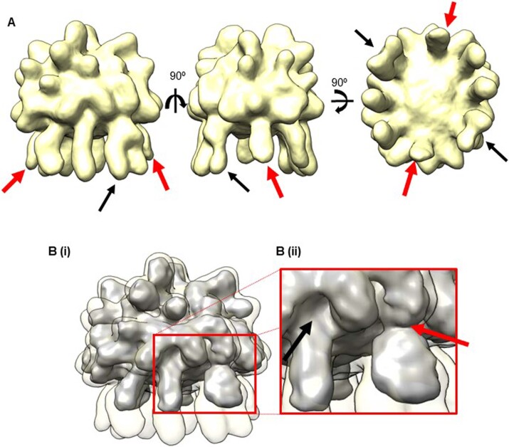
Extended Data Fig. 3. Fibrillin arm region reconstruction.
a) 3D reconstruction of the bead region shown in three orthogonal orientations. There are 8 arms which are visible emerging from the bead structure. Highlighted with black arrows are two arms which are wider. The red arrows highlight two arm regions which are masked out in the final bead reconstruction. b) The reconstruction in (A) is shown overlaid on the final bead reconstruction shown in grey. Panel (Bii) shows a magnified view of the arm regions connecting to the bead region. The red arrow shows how the wider arms connects to the outside of the bead, in contrast the arm highlighted by the black arrow inserts into the centre of the bead.

