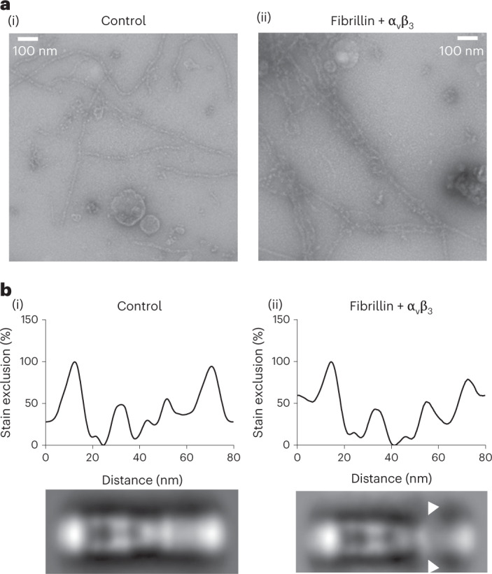
Fig. 6. Integrin αvβ3 headpiece-binding site.

a, Negative stain images of (i) control microfibrils and (ii) microfibrils in complex with the integrin αvβ3 headpiece. b, Stain exclusion plots (top) and class average images (bottom) of (i) 356 control microfibril periods and (ii) 391 microfibril periods complexed with the integrin αvβ3 headpiece. The arrowheads indicate a region at the interbead/shoulder junction with less well-defined structure and diffuse stain exclusion in the presence of the integrin αvβ3 headpiece. The aligned and averaged microfibril periods from two biological repeats are shown.
