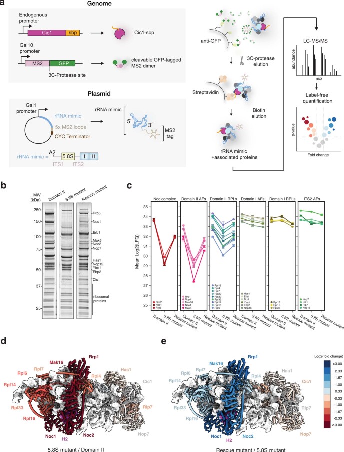
Extended Data Fig. 10. Disruption of helix 2 prevents assembly of domain II of the 25S rRNA.
(a) Schematic depiction of two-step purification of Domain II mimics followed by mass-spectrometry analysis. Purification of the mimics has been performed similarly to purification of Noc1 RNP except endogenous Cic1 has been C-terminally tagged with streptavidin-biding peptide (sbp). Following purification, samples were analyzed by liquid chromatography-tandem mass spectrometry (LC-MS/MS) and obtained data normalized using label-free quantification. (b) SDS-PAGE analysis of purified Domain II rRNA mimics. (c) Changes in mean protein abundance, expressed as LFQ values, between purified rRNA mimics: Domain II, 5.8S mutant disrupting helix 2, and rescue mutant restoring helix formation. Error bars represent SD of 3 replicates. (d, e) Structure of Noc1-Noc2 RNP colored accordingly to log2 fold change in protein abundance between (d) 5.8S mutant and Domain II and (e) rescue mutant and 5.8S mutant. Proteins are depicted as cartoons and rRNA as white surface. Helix 2 (H2) is shown in magenta.

