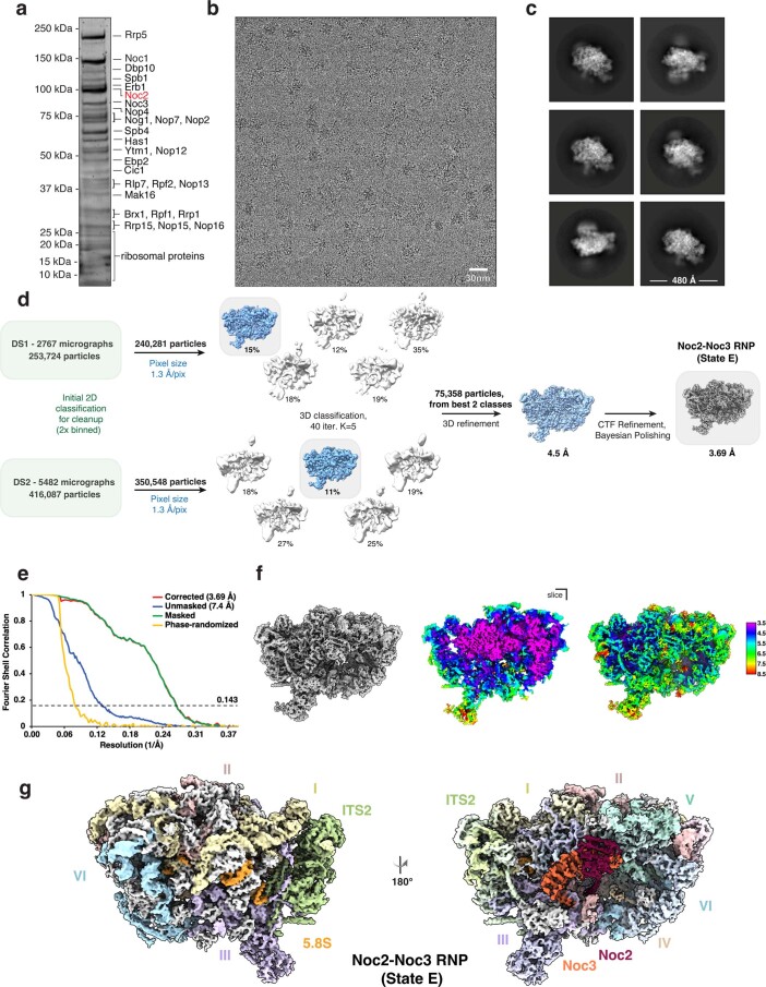
Extended Data Fig. 2. Purification and structure determination of the Noc2-Noc3 RNP (State E).
(a) Representative SYPRO Ruby stained SDS-PAGE of purified fraction containing the Noc2-Noc3 RNP (State E) pre-ribosomal particles. Molecular weight markers are indicated on the left (MW) and bait protein (Noc2; red) and other bands were identified by LC-MS/MS analysis of the eluate. Protein gel (and purifications) were repeated multiple times (n>3) with similar results. (b) Representative motion-corrected cryo-EM micrograph of the Noc2-Noc3 RNP particle sample. (c) Six most populated 2D class averages (1.3 Å/px), box size 440 pixels, mask diameter 480 Å. (d) Cryo-EM data acquisition and processing workflow to obtain the final reconstruction of the Noc2-Noc3 RNP at a resolution of 3.69 Å. (e) Solvent-corrected FSC curves for the Noc2-Noc3 RNP, calculated in Relion 3.0, with reported resolutions determined at FSC=0.143 upon post-processing. (f) Local resolution estimation displayed in rainbow color on the cryo-EM map of the Noc2-Noc3 RNP. (g) Final cryo-EM reconstruction of the Noc2-Noc3 RNP with rRNA domains, assembly factors and ribosomal proteins (in gray) identified and colored.

