
Figure 1. Proband skeletal imaging confirms urate crystal deposition in multiple joints and erosive disease.
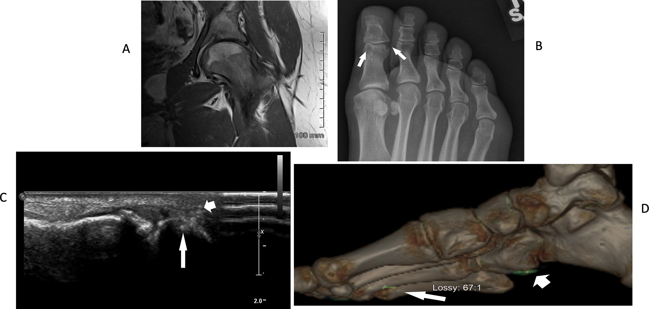
A. Left hip MRI revealed inflammatory synovitis, joint effusion, and articular cartilage loss. B. Foot radiographs revealed classic punched out great toe joint interphalangeal joint erosions, with well-defined margins (arrows), features characteristic of tophaceous, erosive gout. C. First MTP joint ultrasound showed hyperechoic areas consistent with urate crystal deposits (arrowhead), accompanied by cortical erosion (arrow). D. Dual energy CT exams of the foot confirmed urate crystalline macroaggregates affecting foot joints and surrounding soft tissues (arrow and arrowhead).