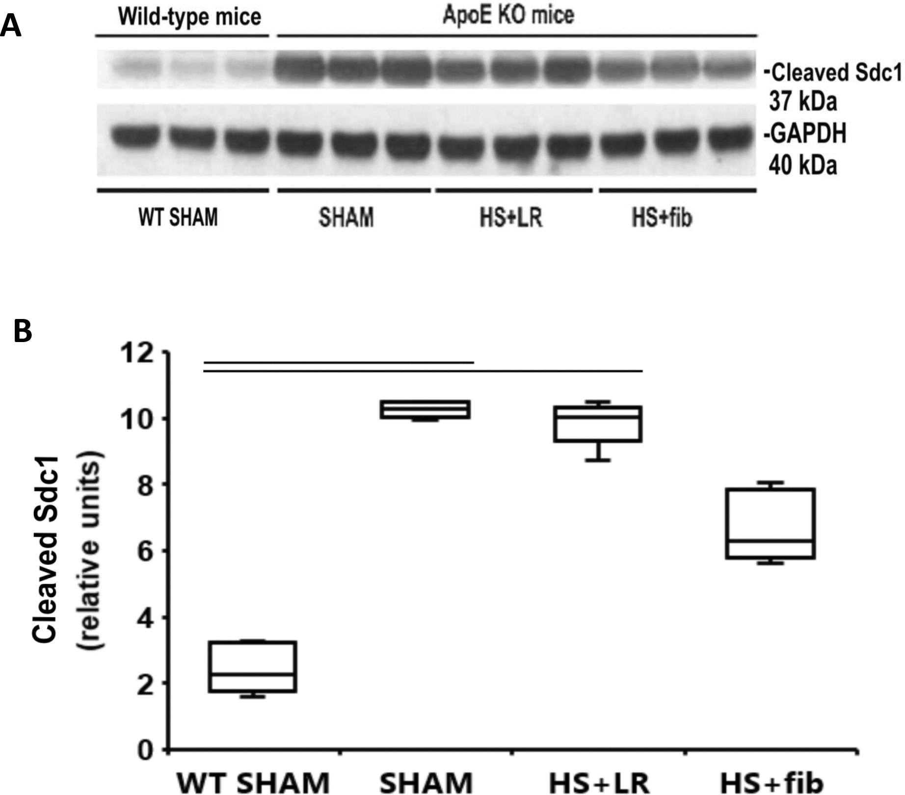
Fig. 5.

Western blot analysis of cleaved syndecan-1 fragments in the lung tissues. Mice were treated as described in Fig. 1. Lung tissues were homogenized for electrophoresis and Western blot analysis. A: Representative immunoblotting for cleaved syndecan-1 fragment and loading control GAPDH. B: Summaries of active MMP-9 band intensities (normalized to GAPDH). The values are presented as medians with IQR. Bars indicate relationships with p<0.05; n=5 per group.
