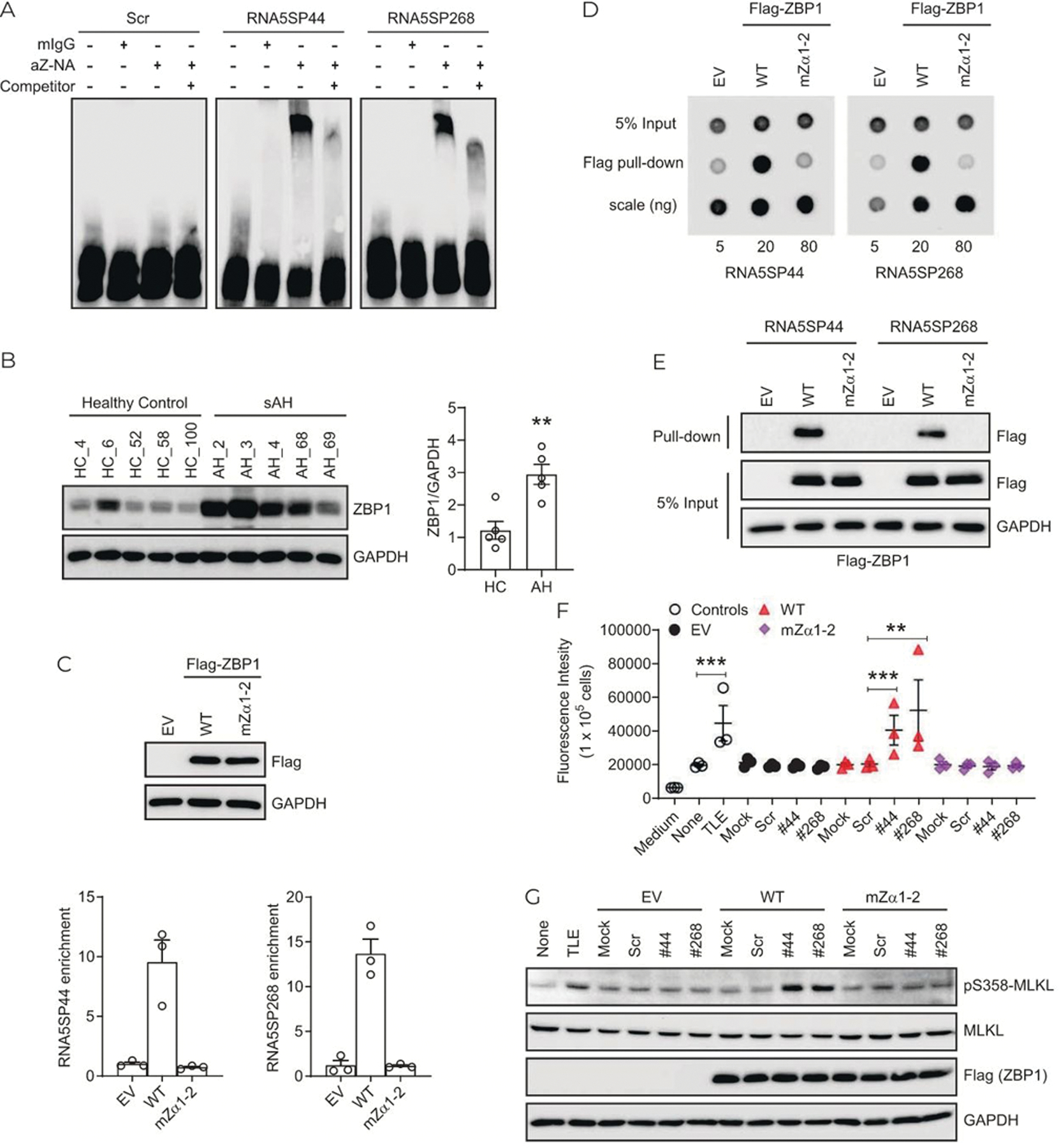
FIGURE 7.

RNA5SP transcripts contribute to ZBP1-mediated cell death. (A) REMSA assay. A 3′-end biotinylated in vitro-transcribed RNA5SP transcripts were incubated with the Z-NAs antibody or a mouse IgG (mIgG) isotype control. A 160-fold nonbiotinylated RNA5SP transcripts (competitor) were used to compete for the binding. (B) Western blot (left) and the quantification using ImageJ (right). **p < 0.01. (C) RNA immunoprecipitation to detect the interaction between endogenous RNA5SP transcripts and ZBP1 in THP-1 cells overexpressing. Empty vector (EV), flag-tagged wild-type ZBP1 (WT), or Zα-domain-mutated ZBP1 (mZα1–2). Top: Western blot of ZBP1 expression. Bottom: The enriched RNAs in the immunoprecipitation were quantified in quantitative PCR and normalized to EV. Data are mean ± SEM of 3 independent experiments. (D) Dot blot to examine the binding of immunopurified flag-ZBP1 or its Zα-domain mutant with 3′-end biotinylated in vitro-transcribed RNA5SP RNAs. The detection was performed with streptavidin antibody against RNAs isolated from anti-flag pull-down. (E) RNA pull-down assays as described in Figure 5E. (F) Cell death was measured by the incorporation of cell impermeant SYTOX Green (488/523 nm). THP-1 cells (1 × 10⁵/well) were transfected with 20 ng of EV or ZBP1, along with 50 fmol of scrambled or RNA5SP RNAs in 96-well plates for 24 hours, and then treated with emricasan (5 μM) for 16 hours. TLE (TNF, 50 ng/mL; LCL-161, 5 μM; emricasan, 5 μM; 16 hours) was used as a positive control. Data are mean ± SEM of 3 independent experiments, **p < 0.01, ***p < 0.001. (G) Western blot using whole cell lysates of THP-1 cells as treated in (F). Abbreviations: RNA5SP, 5S rRNA pseudogene; sAH, severe alcohol-associated hepatitis.
