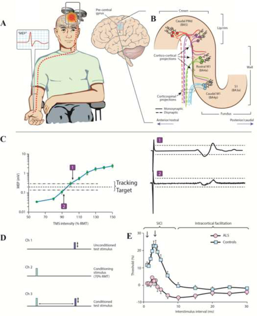
Figure 1: Principles of single and paired-pulse TMS.
(A) Transcranial magnetic stimulation using a figure of eight coil and applied over the primary motor cortex (M1), elicits a motor evoked potential (MEP, red potential in inset) from a target muscle. (B) Candidate descending corticomotoneuronal pathways from the precentral gyrus that contribute to the MEP response. Direct neuronal activation most likely occurs in the lip/rim regions of the motor hand knob. Activation spreads to the rostral and caudal parts of the M1, via cortico-cortical synaptic transmission, potentially contributing to indirect waves (I-waves). There is a greater preponderance of fast-conducting monosynaptic cortico-motoneuronal neurons in the caudal M1 (BA4p) compared to the rostral M1 (BA4a) is highlighted. The exact transition between rostral M1 and caudal dorsal premotor cortex (PMd) in the lip/rim region of the gyrus is gradual and varies across subjects. Additional corticospinal pathways may be activated by TMS via excitation of postcentral primary somatosensory cortex (S1) and its cortico-cortical projections to rostral/caudal M1. (C) For threshold tracking TMS, a target of 0.2 mV (±20%) is selected which lies in the steepest portion of the stimulus response curve. As such, if the MEP response is larger than the tracking target (potential-1) the subsequent stimulus intensity is reduced, while if the MEP response is smaller than the tracking target (potential-2), the subsequent stimulus intensity is reduced. (D) The paired pulse paradigm is illustrated. Channel 1 records an unconditioned test stimulus, defined as TMS intensity required to generate and maintain the tracking target, which signifies the resting motor threshold (RMT) when using the threshold tracking technique. Channel 2 monitors the subthreshold conditioning stimulus (does not generate MEP) and channel 3 records the conditioned-test stimulus at interstimulus intervals of 1–30 ms. (E) When utilizing the threshold tracking TMS technique, short interval intracortical inhibition (SICI) is represented as increased conditioned-test stimulus intensity required to generate and maintain the tracking target, developed between 1–7 ms. Intracortical facilitation is represented as reduced conditioned-test stimulus intensity. In amyotrophic lateral sclerosis (ALS) patients SICI is reduced and ICF increased, signifying cortical hyperexcitability.

