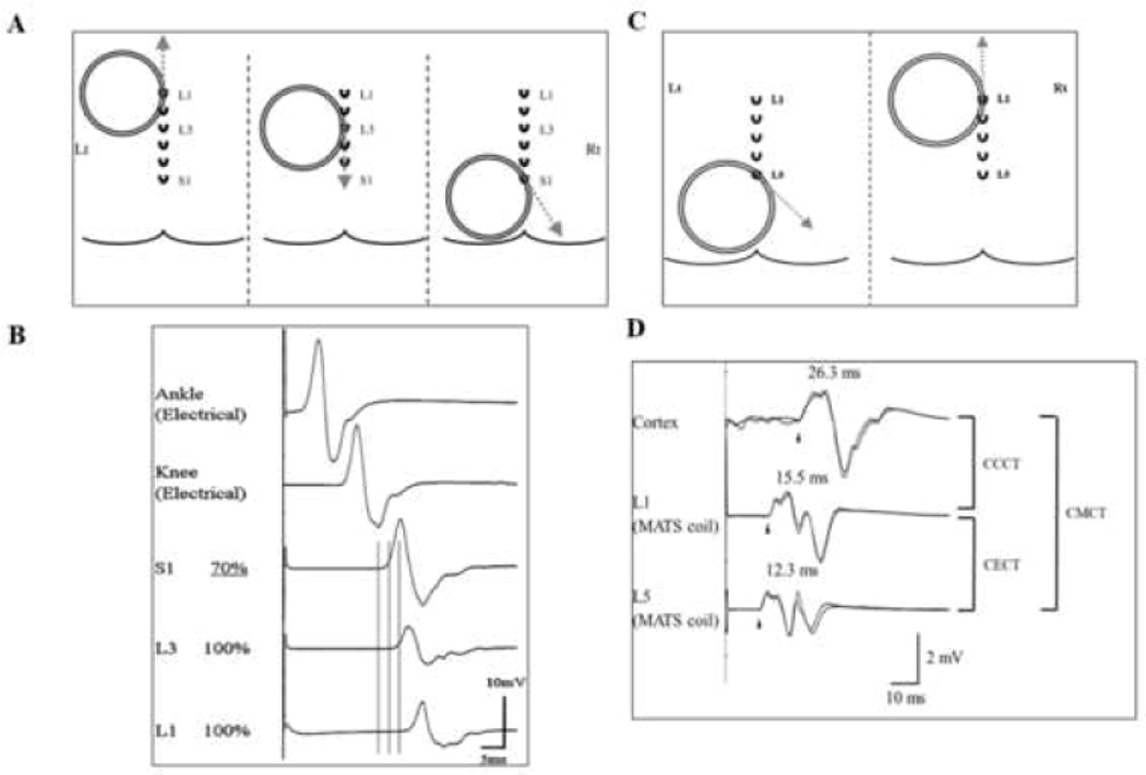
Figure 3: Stimulation of the lumbosacral region.
(A) Position of the magnetic augmented translumbosacral stimulation (MATS) coil in magnetic stimulation of cauda equina with motor evoked potentials (MEP) recorded over the adductor hallucis (AH). The coil edge was positioned over the L1, L3, and S1 spinous processes. The induced current directions are illustrated by grey dashed lines and are tangential to the direction of coil winding over the activation sites. (B) At L1 and L3 levels, MATS coil stimulations failed to elicit a supramaximal MEP response. At the S1 level, stimulating nerves within the neuro-foramina, the MEP responses are supramaximal elicited at a TMS intensity of 70% maximal stimulator output. The MEP onset latency differences between the L1 and L3 stimulation levels suggest that cauda equina in the spinal canal at L1 and L3 levels were activated separately. Cauda equina conduction time (CECT) is calculated by subtracting the S1 from L1 elicited MEP onset latencies when recoding from AH. Tibial nerve compound muscle action potential (CMAP) responses were illustrated with ankle and knee stimulation. (C) Conus stimulation method when recoding over the right tibialis anterior muscle. For proximal cauda equina stimulation, the edge of MATS coil is positioned over the L1 spinous process for inducing currents in an upward direction (dashed grey arrow), while for neuroforaminal activation the edge of the MATS coil is positioned over L5 with induced current direction being 45° downward from a horizontal direction. (D) The MEP responses elicited with cortical, L1 and L5 stimulation are illustrated. The cortico-conus motor conduction time (CCCT) is calculated by subtracting the MEP onset latency elicited by L1 from cortical stimulation. Additionally, CECT is measured by subtracting MEP onset latency elicited by L1 form L5 stimulation. Central motor conduction time (CMCT) is represented by addition of CCCT and CECT.

