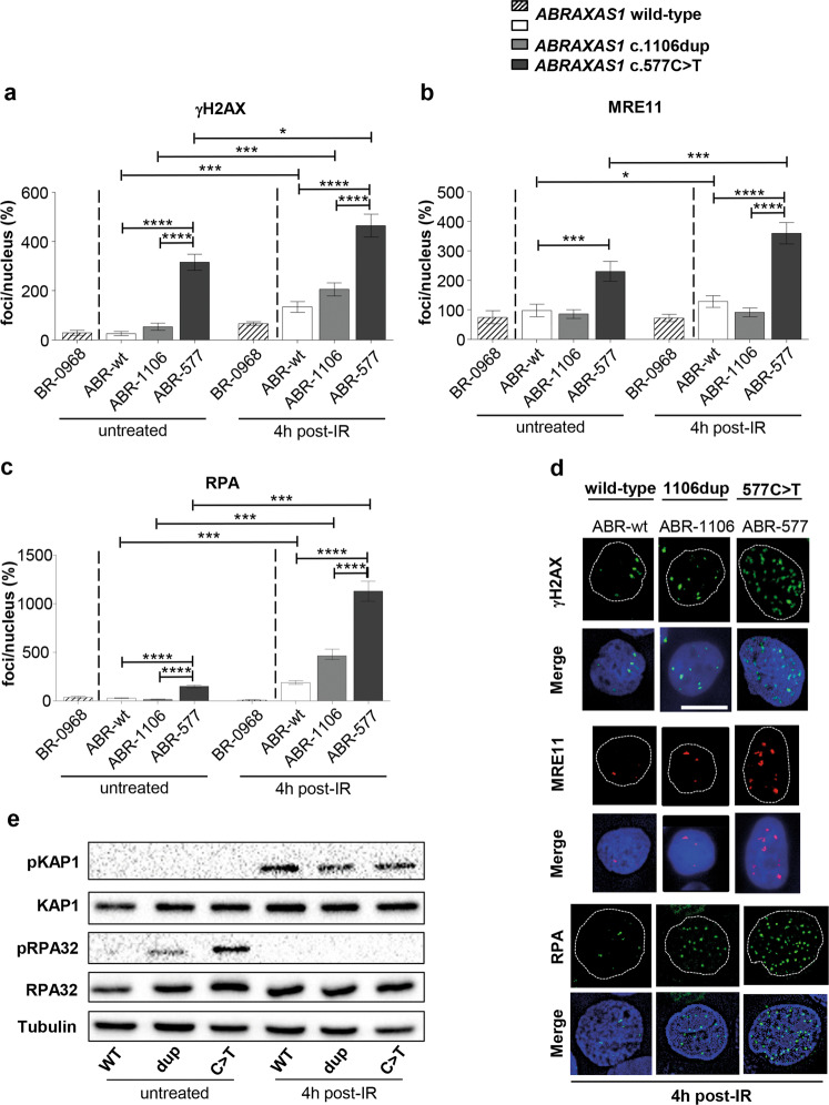
Fig. 4. Monitoring replication stress and DSB responses.
Wild-type ABRAXAS1 (ABR-wt: white and external control BR-0968: hatched), ABRAXAS1 c.1106dup (ABR-1106: light grey) and ABRAXAS1 c.577C>T (ABR-577: dark grey) LCLs were IR-treated and re-cultivated for 4 h (a–d). Foci per nucleus were scored by automated quantification and normalized to the mean foci numbers per nucleus calculated from irradiated wild-type LCLs (ABR-wt and external control BR-0968) measured on the same day (100%). Calculation of statistically significant differences between mean values in the LCLs ABR-wt, ABR-1106 and ABR-577 via Kruskal–Wallis test followed by two-tailed Mann–Whitney U test. Columns show mean values; bars, SEM; *P < 0.05, ***P < 0.001, ****P < 0.0001. a γH2AX-foci per nucleus (n = 150–200 from three independent experiments; absolute values corresponding to 100%: 7.2). (b) MRE11-foci per nucleus (n = 100 from two independent experiments; absolute values corresponding to 100%: 1.9). c RPA-foci per nucleus (n = 100 from two independent experiments; absolute values corresponding to 100%: 10.3). d Representative images of foci (γH2AX: green, MRE11: red, RPA: green) in DAPI-stained nuclei (blue). e Analysis of RPA32 and KAP1 phosphorylation. Representative western blot (n = 2) showing immunodetection of KAP1 phosphorylated on Ser824, RPA32 on Ser33 and total KAP1 as well as RPA32 in control LCL transfected with expression plasmids for wild-type (wt) or mutant ABRAXAS1 variants (dup, ABRAXAS1 c.1106dup; C>T, ABRAXAS1 c.577C>T) followed by 48 h of unperturbed growth (untreated) or 24 h of growth, IR and recultivation for 4 h (4 h post IR). Detection of Tubulin served as loading control. Uncropped western blots are shown in Extended Figure E2.

