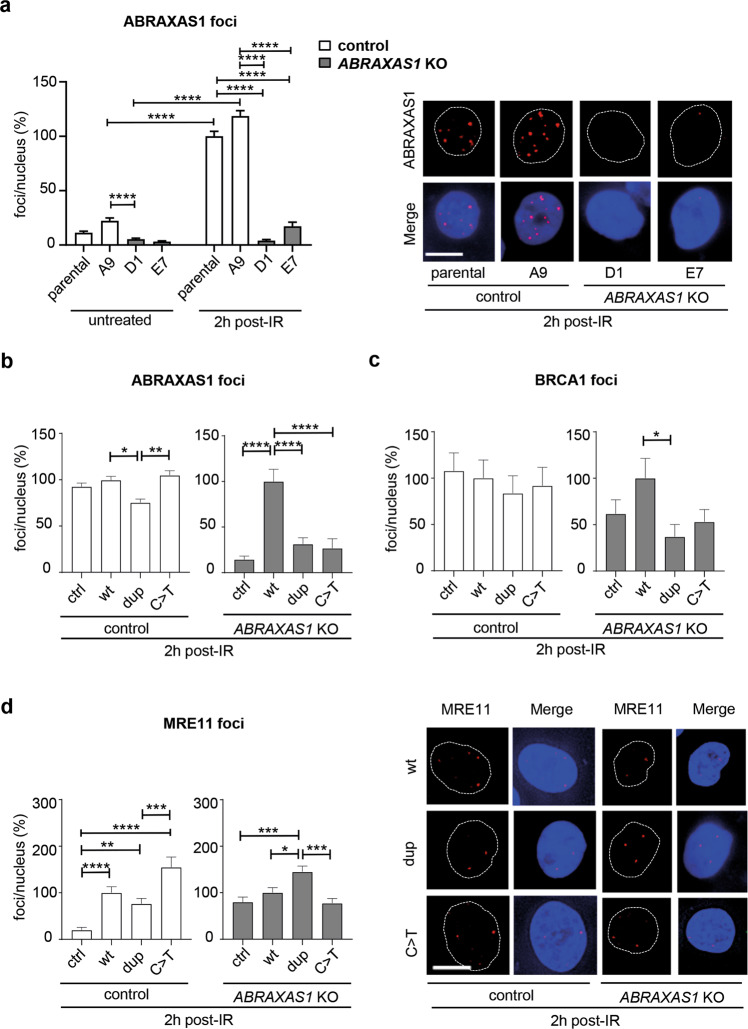
Fig. 6. DDR in mammary epithelial cells as a function of ABRAXAS1.
Mammary epithelial control MCF10A cells (parental and clone A9) and ABRAXAS1 KO cells (clones D1 and E7) were nucleofected with expression plasmids for wild-type ABRAXAS1 (wt), the mutated variants (dup, ABRAXAS1 c.1106dup; C>T, ABRAXAS1 c.577C>T) or empty vector (ctrl), irradiated 24 h later with a dose of 2 Gy and re-cultivated for another 2 h. Nuclear foci were scored and normalized to mean foci numbers of controls on the same experimental day (100%). Columns show mean values (n = 100–400 from two to four independent experiments); bars, SEM; Kruskal–Wallis test followed by two-tailed Mann–Whitney U test; *P < 0.05, **P < 0.01, ***P < 0.001, ****P < 0.0001. a ABRAXAS1-foci per nucleus in the different cell types with and without IR (absolute values corresponding to 100%: 8.9 in irradiated parental MCF10A cells). b–d Foci per nucleus in control (A9) and ABRAXAS1 KO cells (D1) after expression of different ABRAXAS1 proteins and IR-treatment for 2 h. b ABRAXAS1-foci per nucleus (A9 10.3, D1: 3.1 corresponding to 100% in cells expressing wt). c BRCA1-foci per nucleus (A9: 1.2, D1: 0.9 corresponding to 100% in cells expressing wt). d MRE11-foci per nucleus (A9: 1.9, D1: 2.1 corresponding to 100% in cells expressing wt).

