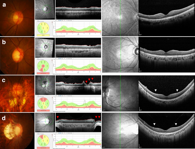
Figure 5.
Representative myopic healthy and glaucomatous eyes. (Left column) Disc photography and (Center column) circumpapillary and (Right column) macular vertical OCT scans. (a) Healthy and (b) glaucomatous eyes without artifacts in circumpapillary OCT scans. (c) Healthy and (d) glaucomatous eyes with large peripapillary atrophy overriding the 12 degree scan circle around the optic nerve for OCT circumpapillary retinal nerve fiber layer scans. Due to image artifacts, segmentation errors were inevitable in circumpapillary OCT scans (red arrowheads). Symmetry and asymmetry are clearly visible (white arrowheads) on macular vertical scans.

