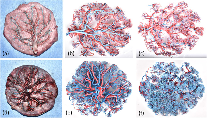
Figure 2.
Visualization of the stages of placental vessel casting. (a, b) Placental microvessels in the GDM group were characterized by a significant reduction in branched vessels, uneven thickness, rigid casting, and dry surfaces, and some branched vessels followed a disordered snake-like course or exhibited reticulate distortion. (c, d) Placental microvessels in the normal group were characterized by a soft and straight course and smooth surfaces, and branches that consistently projected outward to form villous branching vessels.

