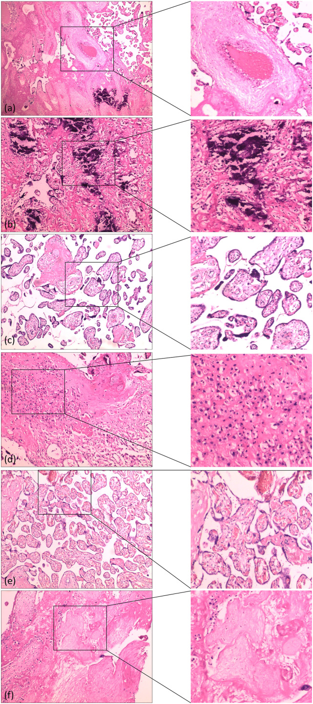
Figure 4.
Photomicrographs of hematoxylin and eosin-stained specimens showing pathologic changes in the placenta. (a) Vessel thrombosis (100× magnification). (b) Villous stroma with diffuse calcifications (200× magnification). (c) Immaturity of chorial villosities with increased villous lumen, with numerous syncytial nodes (200× magnification). (d) Diffuse inflammation (200× magnification). (e) Congestion of villi (200× magnification). (f) Villous fibrinoid necrosis (200× magnification).

