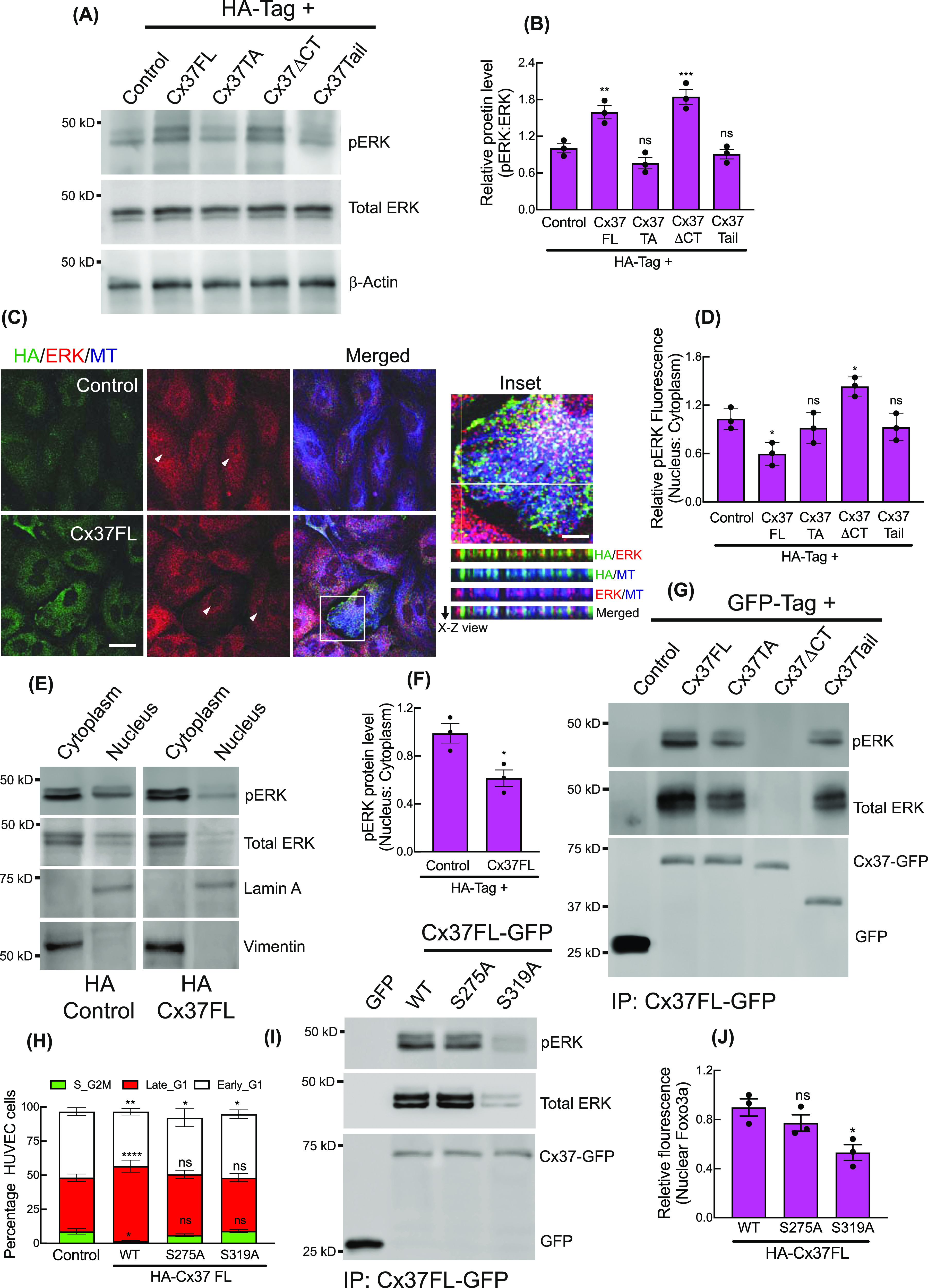
Figure 3. Cytosolic sequestration of Activated ERK by Cx37.

(A, B) ERK and pERK protein levels in Ha-Cx37 construct expressing HUVEC; immunoblot (A) and quantitation (B), Control denoting the expression of HA-Empty vector. (C) Localization of ERK and Cx37 on cytosolic and cortical MT in HA-Cx37FL and HA-Empty vector expressing HUVEC. Inset showing the orthogonal co-localization of Cx37 with ERK and MT along the X–Z axis (white line). Arrowhead indicating the fluorescence of nuclear ERK population in cells. (D) Quantitation of relative fluorescence of pERK in HA-Cx37-expressing cells. (E, F) Relative abundance of pERk protein in nucleus fraction compared with the cytosolic in HA-Cx37FL-expressing HUVEC; immunoblot (E), and quantitation (F). (G) Immunoblot after GFP-trap immunoprecipitation showing the co-immunoprecipitation of ERK and pERK with different Cx37-GFP constructs. (H) HA-Cx37FL-phosphmutants alter cell cycle state of HUVEC-FastFUCCI. (I, J) Immunoblot after GFP-trap immunoprecipitation and quantitation (J) showing Cx37FL-GFP phosphmutants alter ERK and pERK co-immunoprecipitation with Cx37. (B, D, F, H, J) Unpaired t test with Welch correction (F), one-way ANOVA (B, D, J) with Dunnett’s multiple comparisons test, two-way ANOVA (H) with Sidak’s multiple comparisons test. Scale bar: 20 μm ((C); and 5 μm inset).
