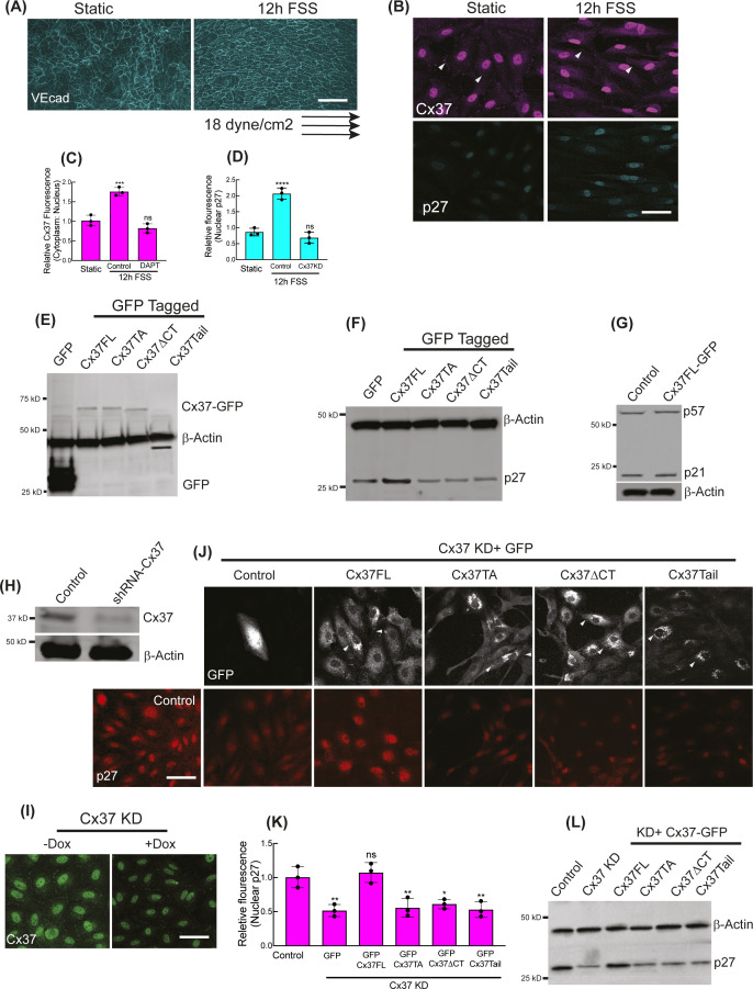
Figure S1. Regulation of p27 expressions by differential Cx37 constructs.
(A, B, C, D) Applied arterial shear stress polarizes HUVEC orientations (A) and increases Cx37 fluorescence intensities (arrowhead showing the Cx37 abundance at cell membrane and cell–cell junctions) and nuclear p27 (B). (C, D) Relative fluorescence quantitations of Cx37 and p27 in response to arterial shear stress are shown here (C, D). Also, the quantitation of the effect of Notch inhibition (by DAPT) on Cx37 fluorescence (C) and the effect of Cx37 knockdown on p27 fluorescence (D) under FSS are shown; respective fluorescence images are not shown. (E) GFP-immunoblot showing the expression of GFP-tagged Cx37 constructs and control GPF-Empty vector (GFP hereafter) in HUVEC. (F) p27-Immunoblot showing the expression of GFP-tagged Cx37 constructs and GFP hereafter in HUVEC. (G) Immunoblots of p27 and p57 in Cx37FL-expressing HUVEC. (H, I) Cx37 immunoblot and Cx37 immunoflorescence (I) are showing the shRNA-mediated and doxycycline-inducible knockdown of Cx37 in HUVEC. (I, J) GFP immunofluorescence of Cx37 constructs and respective p27 fluorescence images and relative p27 fluorescence quantification in p27 knockdown HUVEC. (L) Immunoblot showing the p27 protein level in p27 knockdown HUVEC that expresses Cx37-GFP constructs. (C, D, K) One-way ANOVA (C, D, K) with Dunnett’s multiple comparisons test. Scale bar: 40 μm (B, I, J), and 100 μm (A).

