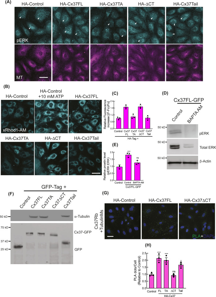
Figure S4. Cytosolic sequestration of activated ERK by Cx37.
(A) Immunofluorescence images showing the pERK localization in HA-Cx37-expressing HUVEC. Arrowhead indicating the pERK fluorescence at nucleus. (B, C) Immunofluorescence of X-Rhod-1 AM Ester indicating the abundance of intracellular Ca2+ in the HA-Cx37-expressing HUVEC; and (C) showing the quantitation of X-Rhod-1 relative fluorescence as a fraction of control; more details are mentioned in methods. (D, E) Intracellular Ca2+ chelation with BAPTA-AM causes inhibition of ERK phosphorylation in Cx37FL-GFP-expressing HUVEC; immunoblot (D) and relative quantitation (E). (F) Immunoblot after GFP-trap immunoprecipitation showing the co-immunoprecipitation of MT with Cx37-GFP constructs. (G) NaveniFlex in-situ rabbit/mouse proximity ligation images showing PLA dots of MT (mouse primary) and Cx37 (rabbit primary) in Cx37-expressing HUVEC; more details are in methods. (H) Quantitation of PLA images of MT-Cx37 in Cx37-expressing HUVEC. 100 cells per conditions, n = 3. One-way ANOVA (C, E, H) with Dunnett’s multiple comparisons test. Scale bar: 20 μm (A) and 40 μm (B, G).

