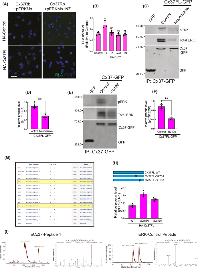
Figure S5. Cytosolic sequestration of Activated ERK by Cx37.
(A, B) NaveniFlex in situ rabbit/mouse proximity ligation images (A) showing PLA dots of pERK (mouse primary) and Cx37 (rabbit primary) in Cx37-expressing HUVEC and quantitation (B). 100 cells per conditions, n = 3. (C, D) MT depolymerization by nocodazole impaired pERK and Cx37 co-immunoprecipitation in Cx37FL-GFP immunoprecipitation by GFP-trap, immunoblot (C), and quantitation (D). (E, F) Inhibition of ERK phosphorylation perturbed ERK-Cx37 co-immunoprecipitation in Cx37FL-GFP immunoprecipitation by GFP-trap, immunoblot (E), and quantitation (F). (G) PhosphoSitePlus-output showing ERK consensus sites on human and mouse Cx37. (H) pERK protein level in Cx37 phosphomutants expressing cells. (I) LC–MS analysis showing ERK-phosphorylated synthetic mCx37 and ERK-substrate peptides in vitro. (B, D, F, H) Unpaired t test with Welch correction (D, F) and one-way ANOVA (B, H) with Dunnett’s multiple comparisons test. Scale bar: 20 μm (A).

