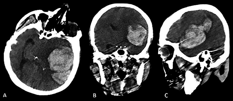
Figure 1. Preoperative Non-Contrast Enhanced Computed Tomography.
A: Preoperative cranial computed tomography axial scan showing a 6x6 cm hyperdense area in the left temporal lobe extending into the intraventricular space, with perilesional edema detected.
B: Preoperative cranial computed tomography coronal scan revealing obliterated major sulci and a faint left lateral ventricle. A mass effect with a 9 mm midline shift was observed.
C: Preoperative cranial computed tomography sagittal scan displaying a 6x6 cm hyperdense area, consistent with hemorrhage and perilesional edema detected.

