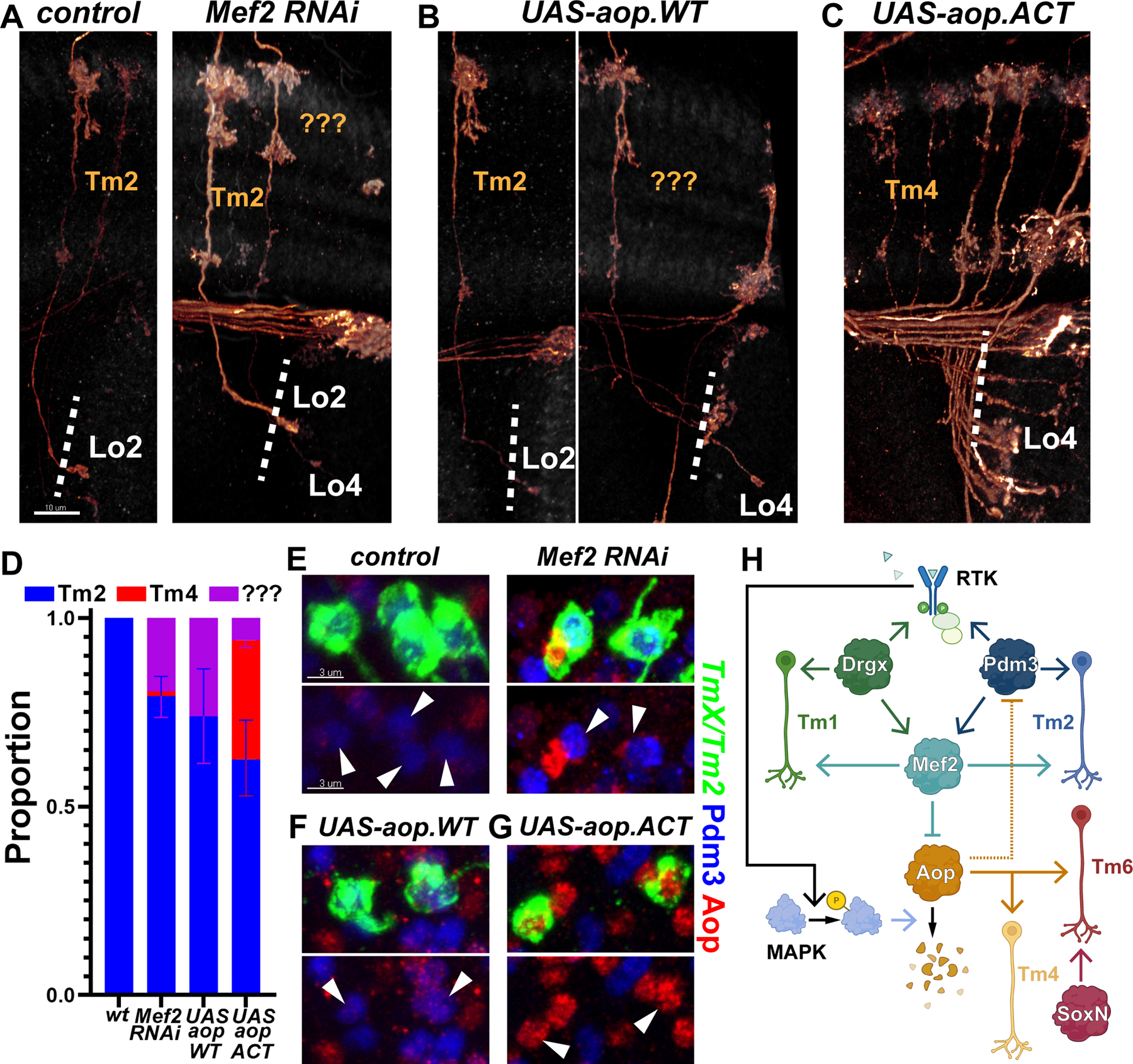
Figure 4: RTK signaling stabilizes the Tm selector network.

A-G, TmX/Tm2-Gal4 driving CD4-tdGFP (flip-out) and Mef2 RNAi (A,E), UAS-aop.WT (B,F) or UAS-aop.ACT (C,G). A-C, 3D reconstructions of GFP for the representative adult neurons in each condition, with anti-NCad. “???” marks neurons that typically target to Lo4 but could not be recognized as any known optic lobe neuron based on their morphology. Dashed lines mark the border of lobula neuropil. D, Quantification of A-C. n= 54/6 (control), 38/8 (Mef2 RNAi), 16/6 (aop.WT) and 75/5 (aop.ACT) neurons/brains. p=0.03 (Mef2 RNAi), p=0.02 (aop.WT), p=0.005 (aop.ACT) for change in cell-type proportions. Error bars denote SEM. E-G, Same as (A-C) with max. projections of somas with anti-Pdm3 (blue) and anti-Aop (red). Arrowheads: GFP+ neurons. Scale bars: 10 μm (A-C) and 3 μm (E-G). H, Summary of the experimentally validated regulatory interactions between Drgx, Pdm3, Mef2 and Aop in Tm neurons. Negative regulation of Pdm3 by Aop (dashed line) is only applicable when Aop cannot be degraded through the MAPK pathway. RTK: receptor tyrosine kinase.
