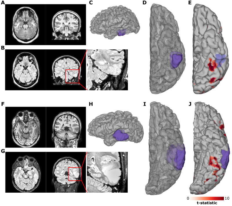
FIG. 2.
A, B, F, and G: Preoperative imaging. Preoperative T1-weighted (A, F) and FLAIR (B, G) MRI for case 1 (A, B) and case 2 (F, G). C, D, H, and I: Lateral (C, H) and ventral (D, I) views of 3D reconstructions of the patients’ cortical surfaces and the FCD (case 1 [C, D]) and tumor (case 2 [H, I]), as determined from the preoperative MRI. E and J: fMRI contrast of words > fixation for case 1 (E) and case 2 (J) with the predicted pial involvement of the relevant pathology highlighted. fMRI thresholded at t > 5.

