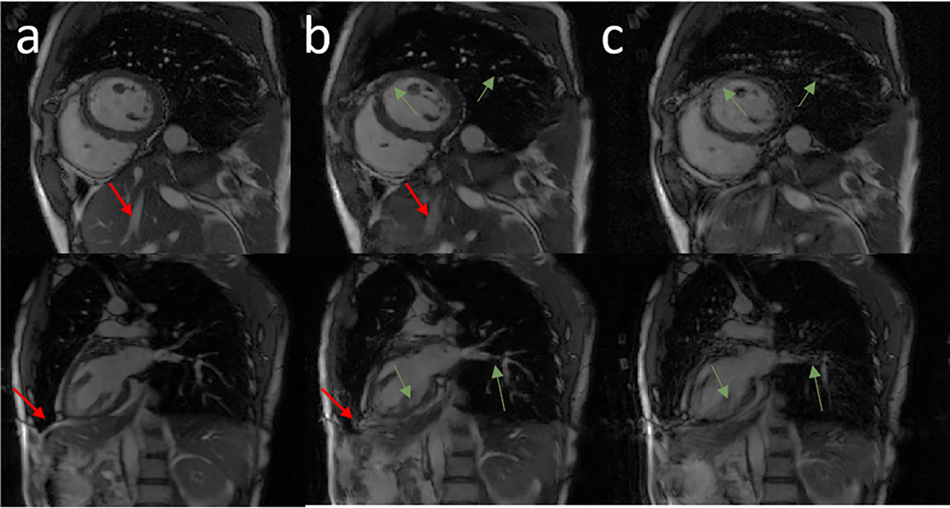
Figure 6.

Representative images in the short-axis view, and vertical long-axis view from two volunteer subjects. Columns a, b, and c show the breath-held cine, motion-corrected free-breathing cine, and motion-corrupted free-breathing cine images, respectively. Green arrows highlight structures that were recovered completely by the network. Red arrows point to regions of residual blurring.
