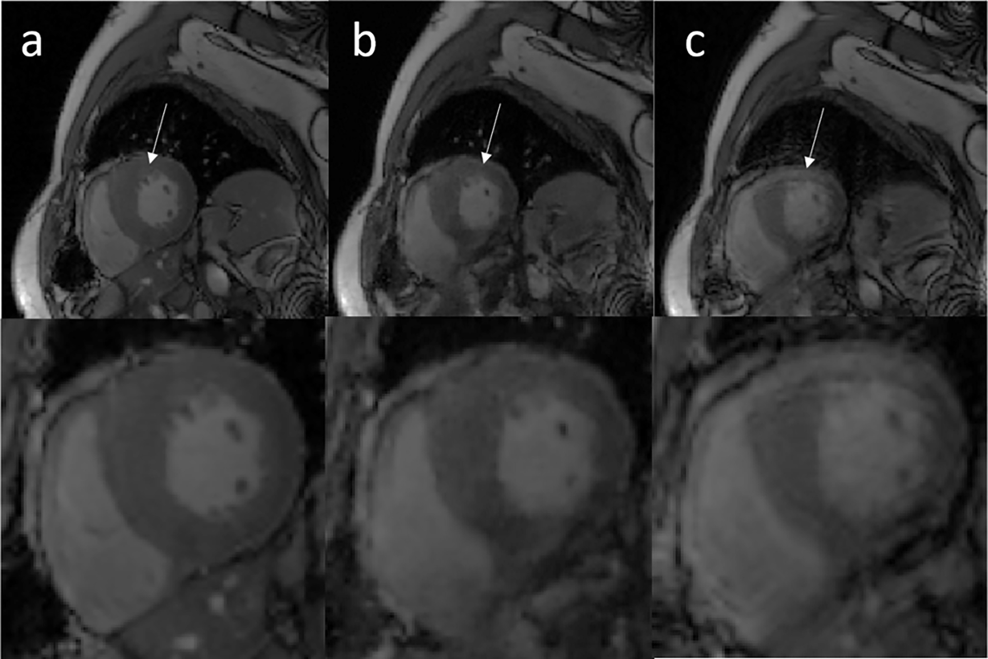
Figure 7.

Representative cardiac cine images from a testing data acquired on a patient. Columns a, b, and c show standard clinical breath-held cine, the motion-corrected cine based on free-breathing data, and motion-corrupted cine data, respectively. White arrows show that the left ventricle region is significantly affected by motion artifacts and these artifacts were removed by the proposed network.
