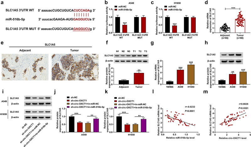
Figure 5.

MiR-516b-5p interacted with SLC1A5 in NSCLC cells. (a) The binding sites between miR-516b-5p and SLC1A5 3ʹUTR WT or MUT was showed. (b-c) The relative luciferase activity of SLC1A5 3ʹUTR WT and MUT reporter vectors was determined by dual-luciferase reporter assay. (d-f) qRT-PCR (n = 66) (d), IHC staining assay (e) and western blotting (f) were used to detect SLC1A5 expression in clinical NSCLC tumor and adjacent normal tissues. (g-h) SLC1A5 expression was measured by qRT-PCR (g) and western blotting (h) in 16HBE, A549 and H1650 cells. (i-k) SLC1A5 protein level was detected by western blotting in A549 and H1650 cells-transfected with sh-NC or sh-circ-OXCT1 and in-miR-NC or in-miR-516b-5p. (l-m) Pearson correlation analysis was performed between relative SLC1A5 level and miR-516b-5p or circ-OXCT1 levels in NSCLC tumor tissues. n = 3 independent biological replicates. Data were showed as mean ± SD. **P < 0.01 and ***P < 0.001 by Mann-Whitney U test, one-ANOVA followed by Tukey’s post hoc test or unpaired t test (two-tailed).
