Fig. 1.

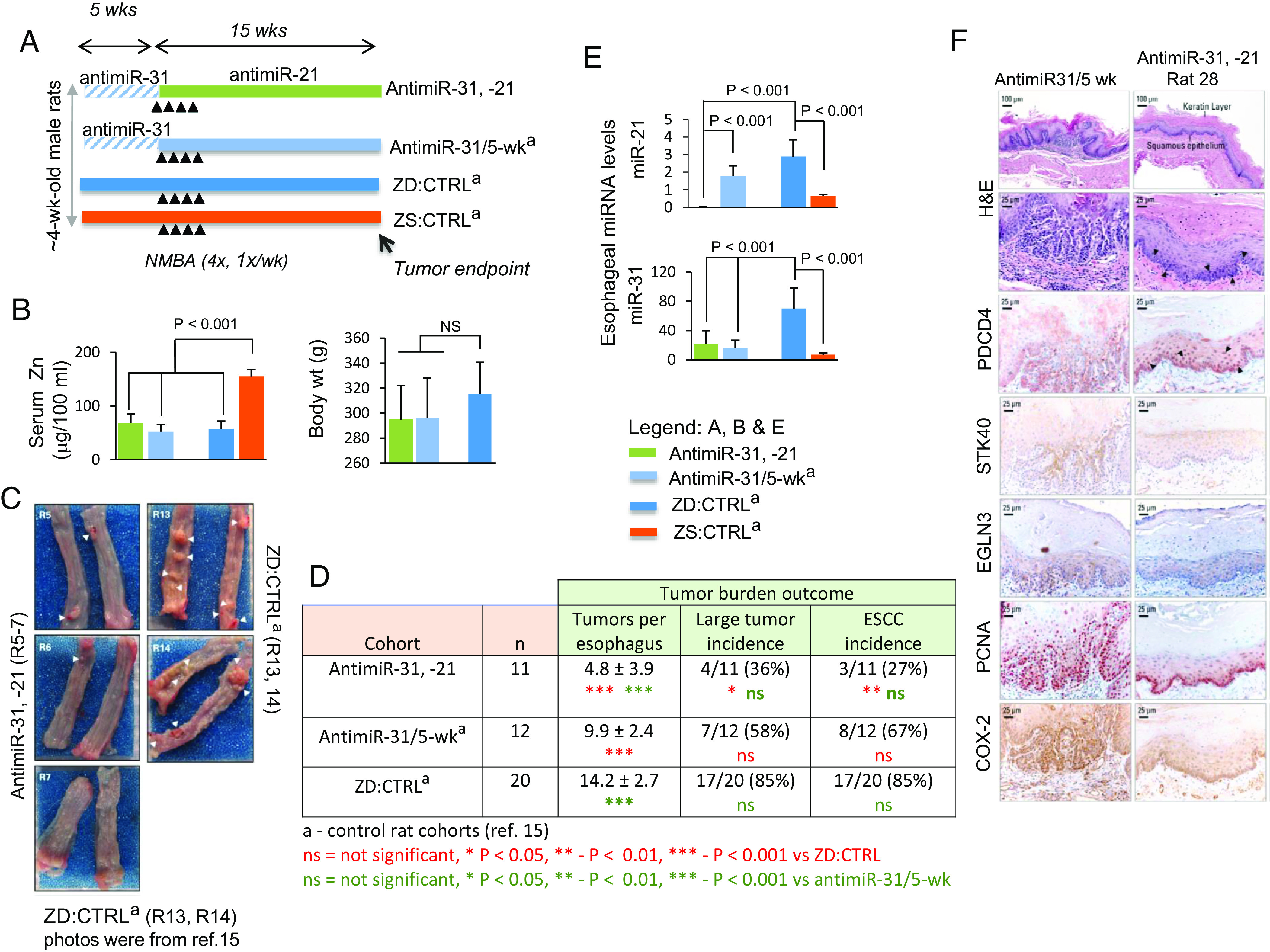
Sequential systemic antimiR-31 delivery followed by antimiR-21 suppresses esophageal carcinogenesis. (A) Study design: this sequential antimiR-31 and antimiR-21 research was done simultaneously with the previous single antimiR-31 delivery study (15) and thus shared the same control rat cohorts as described (15). Over the first 5 wk period, antimiR-31 and antimiR-21 rats (n = 11) were given 10 i.v. doses of antimiR-31. At week 5, the animals received intragastric NMBA doses once a week for 4 wk. Immediately after the first NMBA dose and over 15 wk, antimiR-31 and antimiR-21 rats received their 20 i.v. doses of antimiR-21. The animals were killed 15 wk after first NMBA dose or 48 h after the final antimiR-21 dose. (B) Body weights and serum Zn levels. (C) Macroscopic view of whole esophagus. Representative photos of antimiR-31 and antimiR-21 (R5-7) with isolated/small tumor (arrowheads) vs. representative photos obtained from ZD:CTRLa (R13, R14) (15), showing multiple/large sessile esophageal tumors. (D) Tumor multiplicity (number of tumors/esophagus, mean ± SD; Tukey-HSD (honestly significant difference) post hoc unpaired t test, n = 11 to 20 rats/cohort) (antimiR-31, -21 vs. ZD:CTRL, P < 0.001; antimiR-31 and antimiR-21 vs. antimiR-31/5-wk (P < 0.001); antimiR-31/5wk vs. ZD:CTRL, P < 0.001). Large tumor incidence (size >2 mm), antimiR-31, -21 vs. ZD:CTRL [36 vs. 85%, P < 0.05] and ESCC incidence (%), antimiR-31 and antimiR-21 vs. ZD:CTRL [27 vs. 85%, P = 0.004 (two-tailed Fisher’s Exact test; n = 10 to 20 rats/cohort). (E) qPCR analysis of esophageal miR-31 and miR-21, levels in antimiR-31 and antimiR-21 and control rat cohorts (rat snoRNA as normalizer, Tukey-HSD post hoc unpaired t test. Error bars represent SD; n = 8 rats per group). (F) Sequential delivery of antimiR-31 (5-wk) followed by antimiR-21 (15-wk) induces apoptosis in esophagus antimiR-31 and antimiR-21 (rat 28). Representative photos of antimiR-31/5-wk and antimiR-31 and antimiR-21 (rat 28) showing H&E staining, IHC staining for STK40, EGLN3, & COX-2 (brown, 3,3′-diaminobenzidine tetrahydrochloride, DAB) and PDCD4, & PCNA (red, 3-amino-9-ethylcarbazole substrate-chromogen, AEC). Arrowheads in H&E-stained section (rat 28) indicate the presence of apoptotic cells in esophageal epithelium; arrowheads in near serial PDCD4-stained sections (rat 28) showing abundant/intense nuclear overexpression of PDCD4 (arrowheads), as compared with lack of apoptotic cells (H&E-stained section) and absence of PDCD4 nuclear expression in esophagus of an antimiR-31/5-wk rat.
