Fig. 3.

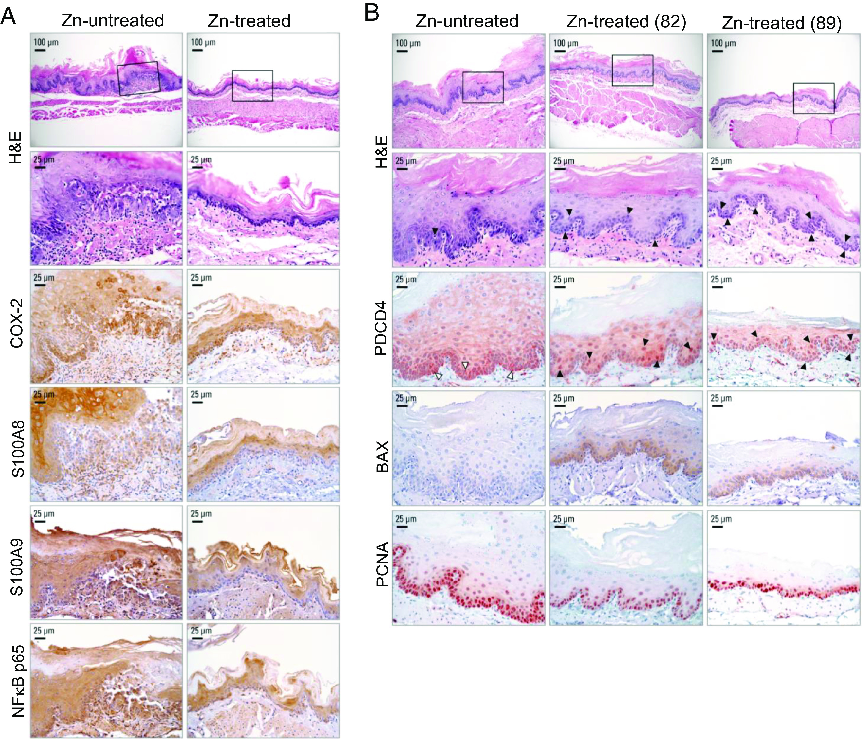
Zn treatment in ESCC-bearing ZD esophagus abolishes inflammation and promotes apoptosis. (A) Zn treatment abolished NF-κB p65-controlled inflammation network. Representative photos of Zn-untreated rats showing strong/abundant IHC staining for all four inflammation markers COX-2, S100A8, S100A9, and NF-κB p65 (brown, 3,3′-diaminobenzidine tetrahydrochloride, DAB) in ESCC bearing hyperplastic esophagus, compared with moderate/diffuse IHC staining for the same inflammation markers in non-proliferative Zn-treated esophagus. (B) Zn treatment triggers apoptosis in Zn-treated esophagus. Black arrowheads in H&E-stained section indicate the presence of apoptotic cells in esophageal epithelium; black arrowheads in near serial PDCD4-IHC stained sections showing abundant/intense nuclear overexpression of PDCD4 in Zn-treated esophagus compared with sporadic occurrence of apoptotic cells (H&E section) and PDCD4 nuclear expression in isolated/sporadic cells of a Zn-untreated rat (white arrowheads). Two esophageal samples from rats 82 and 89 were presented. Representative photos of Zn-treated vs. Zn-untreated esophagus showing H&E staining, IHC staining for PDCD4 (red, 3-amino-9-ethylcarbazole substrate chromogen, AEC), BAX (brown, 3,3′-diaminobenzidine tetrahydrochloride, DAB), and PCNA (red, AEC).
