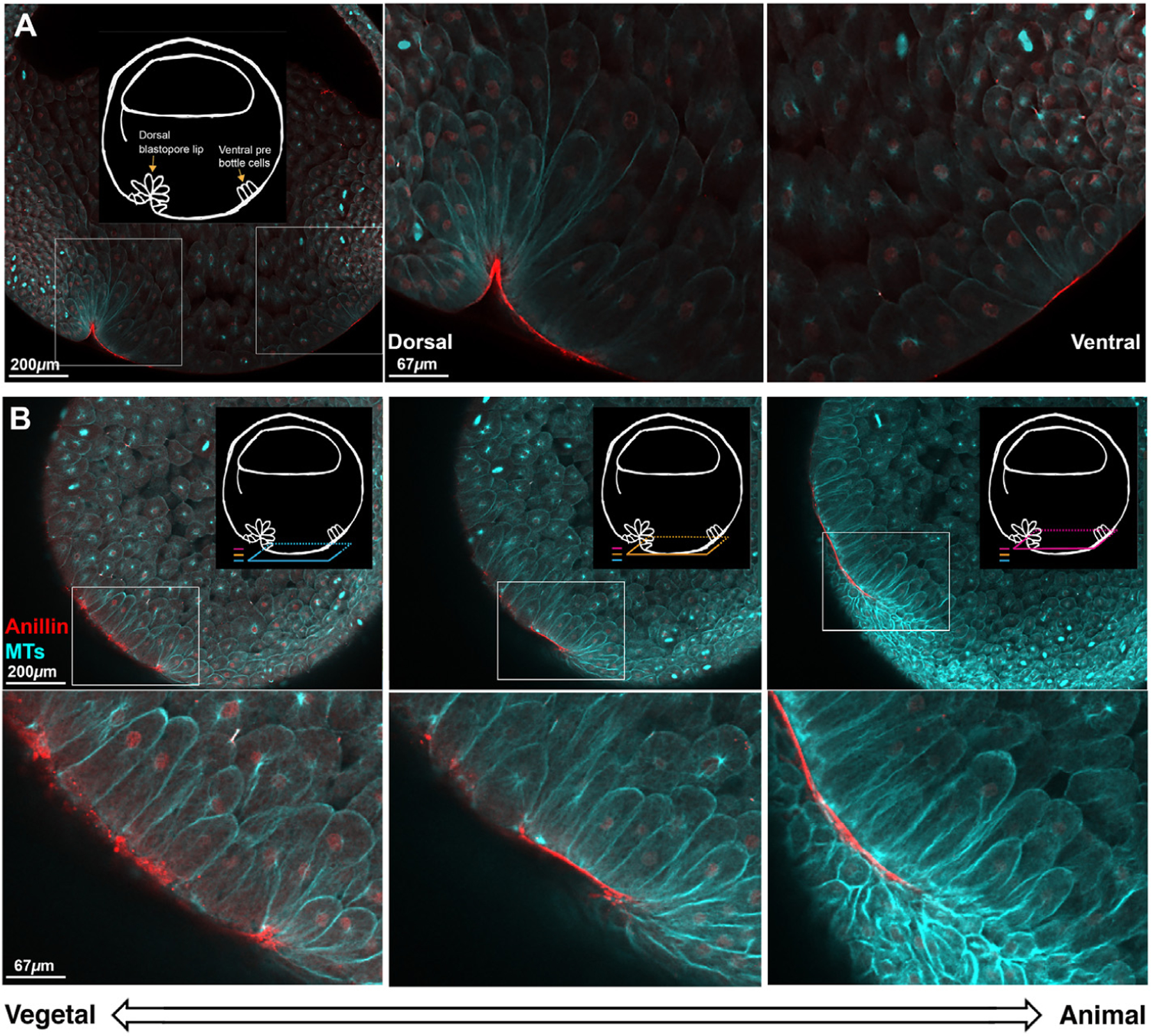
Fig. 5. Whole embryo fixed immunofluorescence against tubulin and anillin during the formation of the early dorsal blastopore lip.

A. Sagittal view of a Stage 10.5 embryo shows membrane proximal anillin (red) localization to dorsal bottle cells and ventral pre-bottle cells. Tubulin (cyan) staining shows the cell outlines revealing bottle cells on the dorsal side of the embryo and elongated cells on the ventral side of the embryo. Schematic highlights the features of interest. Insets show the dorsal and ventral side of the forming blastopore lip at higher magnification. B. Horizontal optical plane views show distinct anillin staining patterns in different parts of the forming dorsal blastopore lip. Three colored planes show the relationship of the horizontal planes to the sagittal plane orientation in A, and the relationship of the horizontal planes to each other. Dorsal is left. Consistent with the sagittal view, anillin has a membrane proximal localization in elongated and apically constricted cells at the dorsal blastopore lip. In the younger, more vegetal region of the lip, the localization of anillin is punctate and in the older, more animal lip, the localization is contiguous and smooth. The z-axis span of the three planes is 90 μ m.
