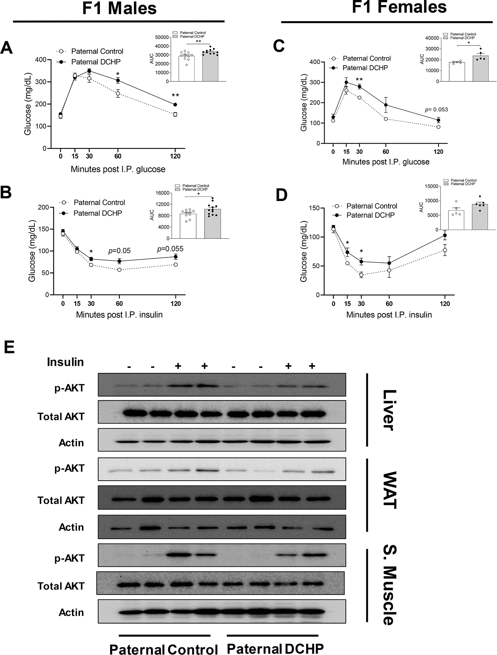
Fig. 3. Paternal DCHP exposure leads to exacerbated diabetic phenotypes and impaired insulin signaling in HFD-fed F1 offspring.

Eight-week-old male WT mice were treated with vehicle control or 10 mg/kg/day of DCHP by oral gavage for 4 weeks. F1 descendants of control or DCHP-exposed sires were fed a HFD for 9 weeks. (A-D) Glucose tolerance test (GTT) and the area under the curve (AUC) of GTT (A and C), and insulin tolerance test (ITT) and AUC of ITT (B and D) in HFD-fed F1 male and female offspring (n = 5–12, two-sample, two tailed Student’s t-test, *P < 0.05 and **P < 0.01). (E) Immunoblotting of phosphorylated Akt and total Akt levels in the liver, white adipose tissue (WAT), and skeletal muscle of F1 male mice injected with saline or 0.35 units/kg body weight insulin (n = 3).
