Figure 3.

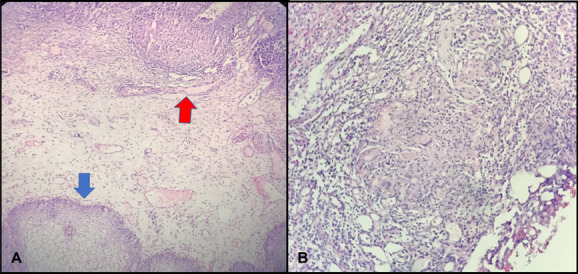
Tur-B pathology specimen image: H&E, ×40 bladder epithelium (blue arrow) and granuloma structure (red arrow) (A); H&E, ×100, granuloma structure in the bladder and Langhans giant cell (B).

Tur-B pathology specimen image: H&E, ×40 bladder epithelium (blue arrow) and granuloma structure (red arrow) (A); H&E, ×100, granuloma structure in the bladder and Langhans giant cell (B).