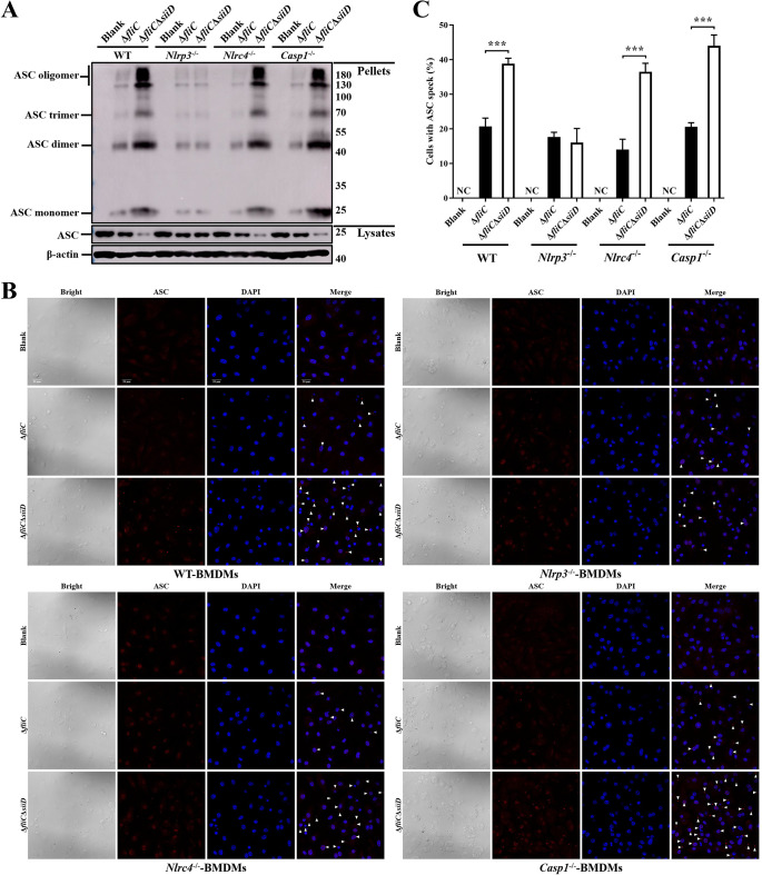
Fig 6. SiiD inhibited the formation of NLRP3 dependent ASC pyroptosome.
BMDMs from WT-, Nlrp3-/—, Nlrc4-/—, and Casp1-/—C57BL/6 mice were primed with LPS (200 ng/mL, 5 h). The Nlrc4-/—BMDMs were infected with ΔfliC or ΔfliCΔsiiD at an MOI of 100:1 for 4.5 h. The WT-, Nlrp3-/—, and Casp1-/—BMDMs were infected with indicated bacteria at an MOI of 50:1 for 4.5 h, uninfected cells were used as a negative control (Blank). (A) Cells were lysed after infection and the pellets were subjected into cross-linking. The ASC oligomerization in the pellets and the total ASC in lysates as the input were examined by western blotting. β-actin was blotted as a loading control. Molecular mass markers in kDa are indicated on the right. (B) The formation of ASC specks (arrowheads) in infected BMDMs were detected by indirect immunofluorescence assay. ASC, red; DAPI, blue. Scale bar, 20 μm. (C) The percentages of cells with ASC speck. Approximately 200 cells were counted in each sample. Data are presented as mean ± SEM of triplicate samples per experimental condition from three independent experiments. ***p < 0.001, as measured by unpaired t test.

