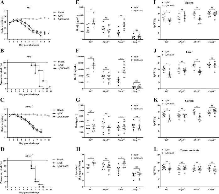
Fig 11. SiiD inhibited NLRP3 inflammasome mediated SE clearance in vivo.
The WT- and Nlrp3-/—C57BL/6 mice were orally infected with ΔfliC or ΔfliCΔsiiD at a dose of 5 × 10⁶ CFU per mouse (n = 10). Uninfected mice were used as the negative control (Blank). The (A, C) body weight changes and (B, D) deaths of mice within 10 dpi were recorded by daily observation. *p < 0.05, as measured by two-way ANOVA for the group differences in weight of mice over time. *p < 0.05, as measured by log-rank (Mantel-Cox) test for the survival curve. The WT-, Nlrp3-/—, Nlrc4-/—, and Casp1-/—C57BL/6 mice were orally infected with ΔfliC or ΔfliCΔsiiD at a dose of 5 × 10⁶ CFU per mouse (n = 6). Serum (E) IL-1β, (F) IL-18, and (G) IL-6 levels in infected mice at 5 dpi were determined by ELISA. (H) Gut inflammation as measured in feces using lipocalin-2 ELISA. Data are presented as the mean ± SEM of log10 ng/g feces. *p < 0.05; NS, not significant, as measured by unpaired t test. The (I) spleen, (J) liver, (K) cecum, and (L) cecum contents homogenates were plated to determine the bacterial CFU per gram of organs at 5 dpi. Data are presented as mean ± SEM of log10 CFU/g. **p < 0.01, ***p < 0.001; NS, not significant, as measured by unpaired t test.

