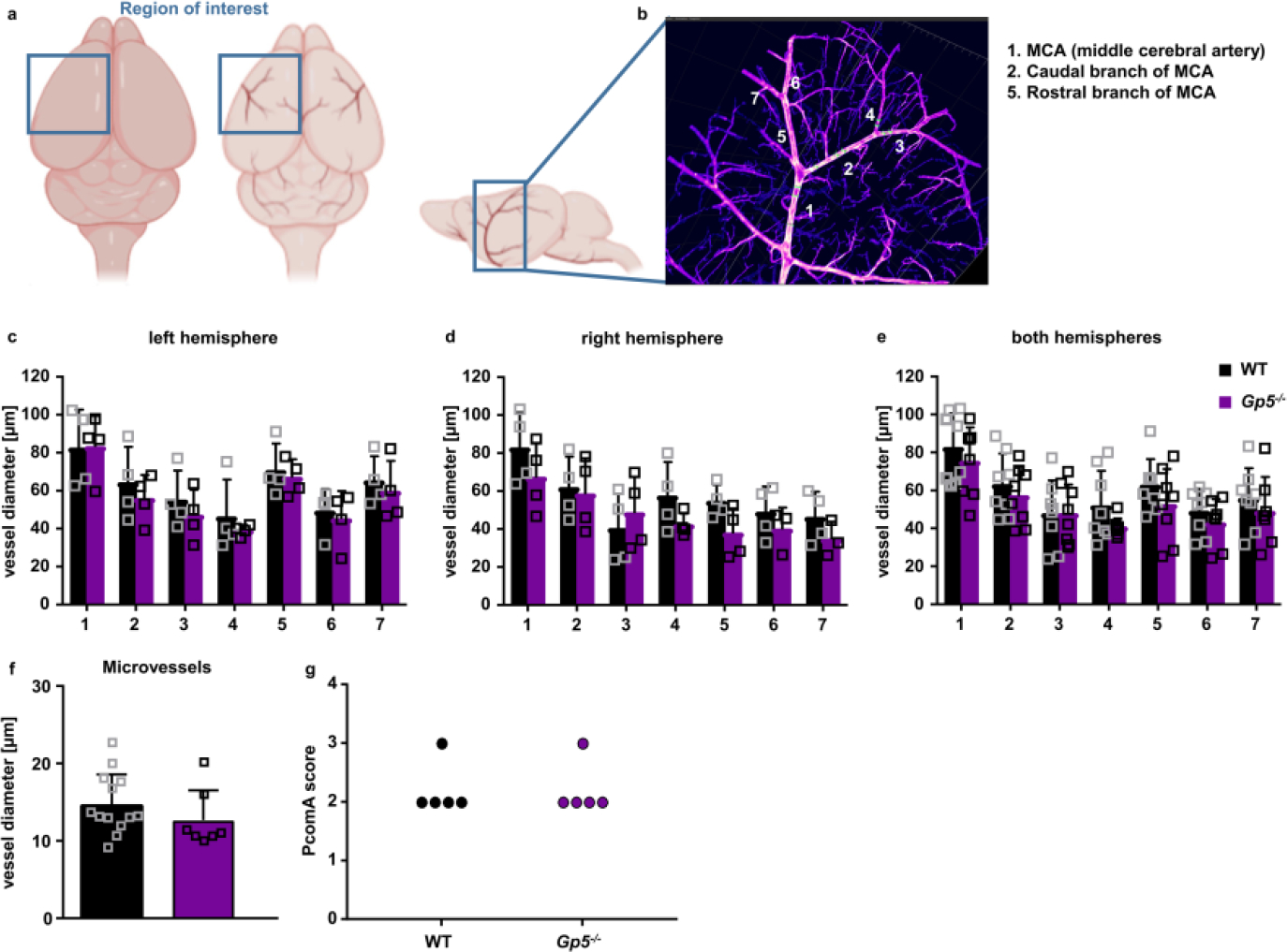
Extended Data Fig. 6. Unaltered MCA vessel diameter in Gp5−/− mice.

Optically transparent brain samples of Gp5−/− and WT mice were imaged using light sheet fluorescence microscopy (LSFM). (a) Due to its conserved branching and its easy recognition, we focused on the region around the middle cerebral artery (MCA) to allow better comparability between the samples. (b) We analysed the vessel diameter of the MCA (1) and 2 subsequent branches of the caudal (2) and rostral (5) branch of the MCA using Imaris Software. (c-e) Vessels in the left, right hemisphere and the combination of both hemispheres did not show any difference between GPV-deficient and WT mice. Mean ± SD. n=4 mice. (f) Vessel diameters of microvessels in the brain were comparable between Gp5−/− and WT mice. Mean ± SD. WT: n=13 vessels of 4 mice, Gp5−/−: n=7 vessels of 4 mice. Two-tailed unpaired t-test with Welch’s correction. (g) PcomA scores (posterior communicating artery) were determined in brains from mice that were perfused with PBS followed 3 ml black ink diluted in 4% PFA (1:5 v/v) and comparable between Gp5−/− and WT mice. n=5. Mann-Whitney test. (a, b) created with BioRender.
