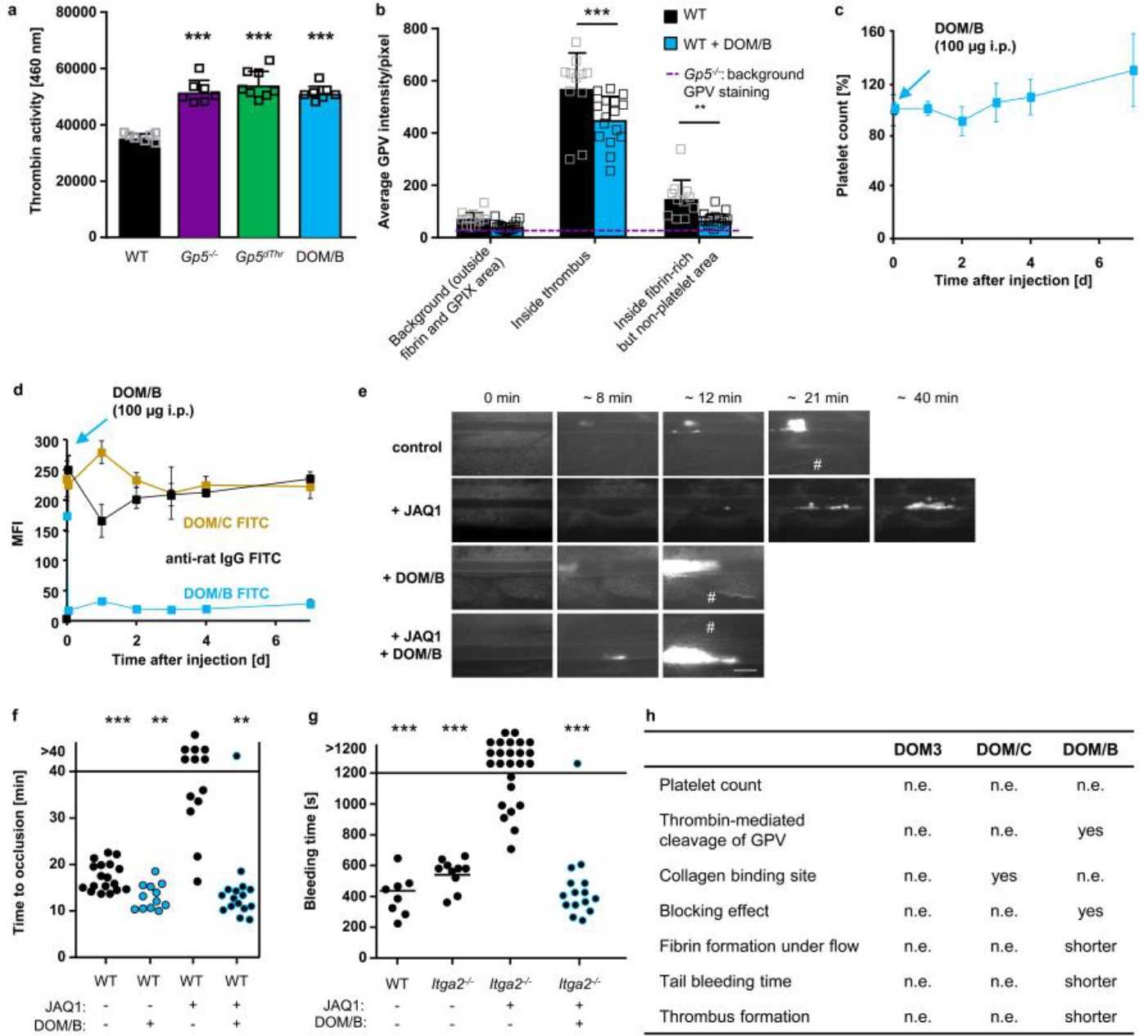
Extended Data Fig. 8. DOM/B restores haemostasis and thrombus formation in the absence of GPVI, thereby reproducing the Gp5−/− phenotype.

(a) Recalcified blood was perfused over collagen/TF spots. Thrombin activity was determined in the outflow using a fluorogenic thrombin substrate and measured immediately. Mean ± SD. WT, Gp5−/−, WT+DOM/B: n=7, Gp5dThr: n=8, one-way ANOVA followed by Tukey’s multiple comparisons test, p<0.0001 for all groups compared to WT. (b) Recalcified blood was perfused over collagen/TF spots. Samples were stained for platelets (anti-GPIX), fibrin(ogen) and GPV, fixed and analysed with a Zeiss Airyscan microscope. Quantification of GPV intensities inside fibrin-rich, non-platelet area. Background GPV intensity in Gp5−/− images is displayed as dashed line (n=4). Mean ± SD. Two-way ANOVA followed by Tukey’s multiple comparisons test, p<0.0001 (inside thrombus), p=0.0089 (inside fibrin), WT. n=11, WT+DOM/B: n=16. (c, d) WT mice were injected with DOM/B (100 μg i.p.) and platelet count was assessed for 7 days (c). Platelet count at d0 was set to 100%. Mean ± SD. n=5 mice, 2 independent experiments. (d) GPV platelet surface expression was assessed by flow cytometry for up to 7 days after DOM/B injection. Mean ± SD. n=5 mice, 2 independent experiments. Receptor opsonisation was measured using an anti-rat IgG FITC antibody. (e-g) GPVI was depleted from the platelet surface by injection of the anti-GPVI mAb JAQ1. Representative images (e) and quantification (f) of thrombus formation upon FeCl3-induced injury of mesenteric arterioles in WT mice after JAQ1 and DOM/B treatment. Thrombus formation in no more than two arterioles of each mouse was analysed; data points represent measurements of one arteriole. WT: n=18 arterioles of 10 mice, WT+DOM/B: n=12 arterioles of 6 mice, WT+JAQ1: n=13 arterioles of 7 mice, WT+DOM/B+JAQ1: n=16 arterioles of 8 mice, two-tailed Fisher’s exact test to compare occluded vs. non-occluded vessels was used. p=0.0007 (vs. WT), p=0.0052 (vs. WT+DOM/B), p=0.0011 (vs. WT+JAQ1+DOM/B), # indicates vessel occlusion. Scale bar: 50 μm. (g) Mice lacking both collagen receptors GPVI and α2 were treated with DOM/B and haemostatic function was assessed using a tail bleeding assay on filter paper. Each symbol represents one mouse. WT: n=8, Itga2−/−: n=10, Itga2−/− + JAQ1: n=25, Itga2−/− + JAQ1 + DOM/B: n=16, two-tailed Fisher’s exact test for open vs. occluded vessels. p=0.0009 (vs. WT), p=0.0003 (vs. Itga2−/−), p=0.0001 (vs. Itga2−/−+JAQ1 +DOM/B), (h) Summary of the effects of the different anti-mGPV antibodies. n.e.: no effect. *p < 0.05; **p < 0.01; ***p < 0.001.
