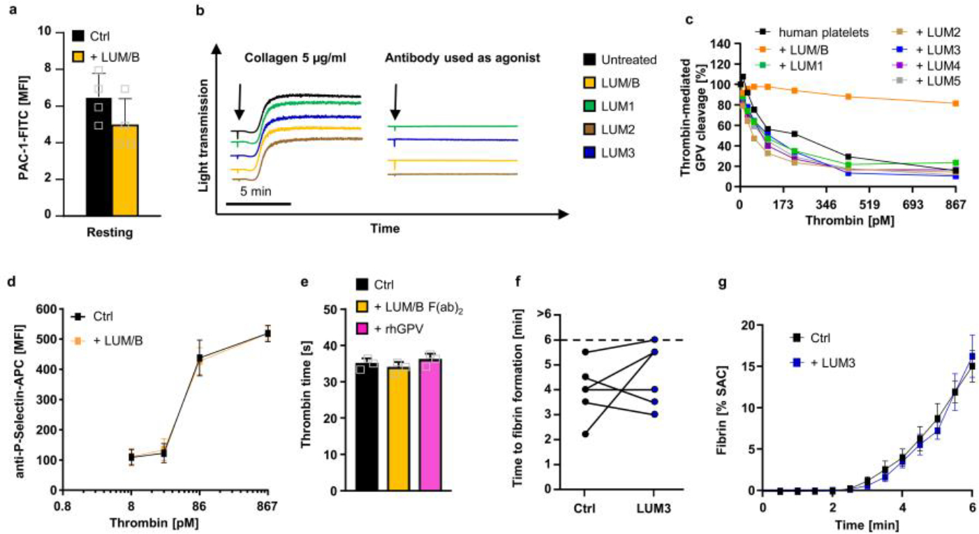
Extended Data Fig. 9. LUM/B has no effect on thrombin-mediated platelet activation.

(a) Human platelets were incubated with 10 μg/ml LUM/B prior to flow cytometric analysis of PAC1-binding. Mean ± SD, n=4 donors. (b) Human platelets were incubated with the indicated anti-hGPV mAbs light transmission was recorded on a Apact four-channel aggregometer over 10 min. Representative curves for n=3. (c) Human platelets were incubated with the indicated antibody (10 μg/ml) prior to thrombin stimulation. Thrombin-mediated cleavage of GPV was assessed by flow cytometry. Mean ± SD, n=2 donors, 3 independent experiments. (d) Flow cytometry reveals unaltered reactivity of LUM/B-treated platelets upon thrombin stimulation compared to controls. (e) Neither blockade of hGPV by LUM/B F(ab)2 nor rhGPV (290 nM) affect thrombin time. (a, c, d, e) Values are displayed as mean ± SD. (d) n=4 donors, 3 independent experiments, two-tailed unpaired t-test with Welch’s correction. (e) n=3 donors, one-way ANOVA. (f, g) Recalcified whole blood was incubated in vitro with 10 μg/ml anti-hGPV antibody LUM3 prior to perfusion over collagen/TF spots. Quantification time to fibrin formation (f) and fibrin surface coverage (g) during blood flow of LUM3-treated (n=8 donors) and control samples (n=10 donors). (f) n=6 donors Values are depicted as mean ± SEM (g). SAC: Surface area coverage. Ctrl: Human donor.
