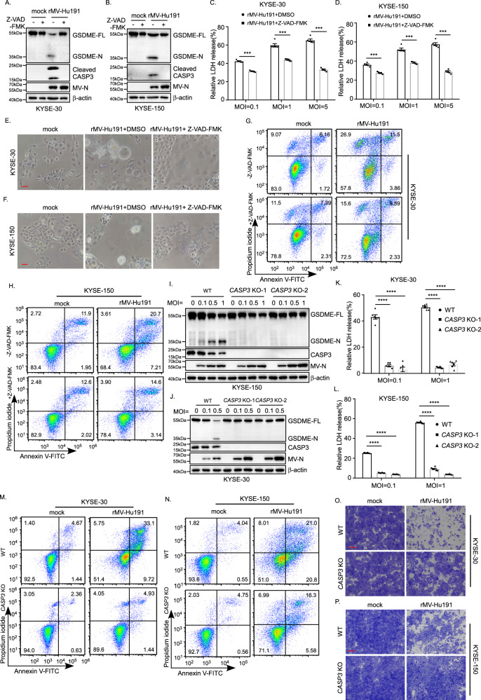
Fig. 4. rMV-Hu191 induces pyroptosis through caspase-3 cleavage of GSDME.
A, B The pan-caspase inhibitor Z-VAD-FMK attenuated GSDME cleavage induced by rMV-Hu191 in KYSE-30 (A) and KYSE-150 (B) cells. Cells were treated by rMV-Hu191 at an MOI of 0.5 with 20 μM Z-VAD-FMK for 48 h and then subjected to immunoblot. C, D Z-VAD-FMK relieved LDH-release cell death induced by rMV-Hu191 in KYSE-30 (C) and KYSE-150 (D) cells. LDH release was measured after rMV-Hu191 treatment for 72 h at the indicated doses with 20 μM Z-VAD-FMK. Data are presented as mean ± SEM (n = 5). ***p < 0.001, two-tailed Student’s t-test. E, F Z-VAD-FMK alleviated the pyroptosis morphology of KYSE-30 (E) and KYSE-150 (F) cells induced by rMV-Hu191. Cells were treated by rMV-Hu191 at an MOI of 0.1 for 48 h, and then the cell morphology was analyzed by an optical microscope. The scale bars represent 50 μm. G, H KYSE-30 and KYSE-150 cells treated by rMV-Hu191 at an MOI of 0.1 with 20 μM Z-VAD-FMK for 48 h were collected and stained with Annexin V-FITC/PI and then subjected to flow cytometry analysis. I, J Deficiency of caspase-3 attenuated GSDME cleavage induced by rMV-Hu191 in KYSE-30 (I) and KYSE-150 (J) cells. Cells were treated by rMV-Hu191 for 48 h at the indicated MOI and then subjected to immunoblot. K, L The LDH release in KYSE-30 (K) or KYSE-150 (L) WT and CASP3 KO cells was measured after rMV-Hu191 treatment for 72 h at the indicated doses. Data are presented as mean ± SEM (n = 5). ***p < 0.001, one-way ANOVA followed by Dunnett’s test. M, N KYSE-30 (M) or KYSE-150 (N) WT and CASP3 KO cells treated by rMV-Hu191 at an MOI of 0.1 for 48 h were collected and stained with Annexin V-FITC/PI and then subjected to flow cytometry analysis. O, P KYSE-30 (O) or KYSE-150 (P) WT and CASP3 KO cells were treated with rMV-Hu191 at an MOI of 0.1 for 72 h. Cell-killing efficiency was then determined by crystal staining. The scale bars represent 500 μm.

