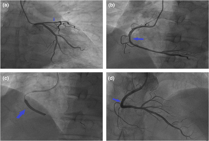
FIGURE 2.

Coronary angiography. (a) The left main trunk was normal, the proximal segment of anterior descending branch was plaque, 30% localized stenosis in the middle segment (arrow), cyclotron branches scattered in plaques. (b) 70% of the RCA was narrowed in the middle and far segment (arrow). (c) A BRS (arrow) was implanted in the middle and distal segments of the RCA. (d) The blood of the RCA resumed to flow smoothly (arrow).
