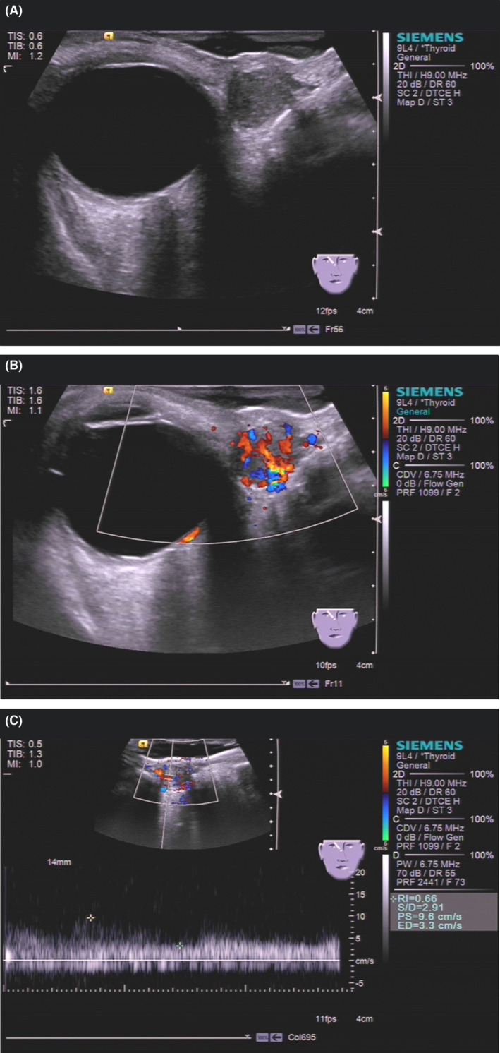
FIGURE 1.

Ultrasonography of superficial angiomyxoma. (A) Grayscale ultrasound showed that the tumor was oval, defined, hypoechoic, homogeneous, and 12 mm × 10 mm in size. (B) Color Doppler flow imaging showed that multiple small and main vessels were visualized inside the tumor. (C) Pulsed‐wave Doppler showed the systolic blood flow velocity was 9.6 cm/s with a high resistance index.
