
FIGURE 2.
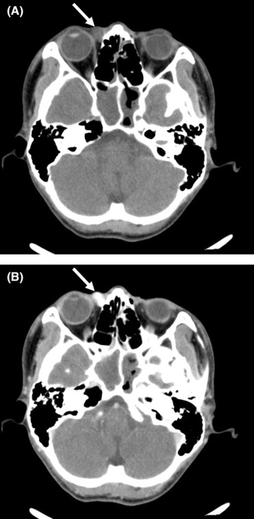
Computed tomography of superficial angiomyxoma. (A) Plain computed tomography showed that the tumor (arrow) under the medial rectus muscle of the right eye was nodular and comparatively defined with a value of 44 HU. (B) Enhanced computed tomography showed that the tumor (arrow) was obviously enhanced with a value of 157 HU–178 HU.