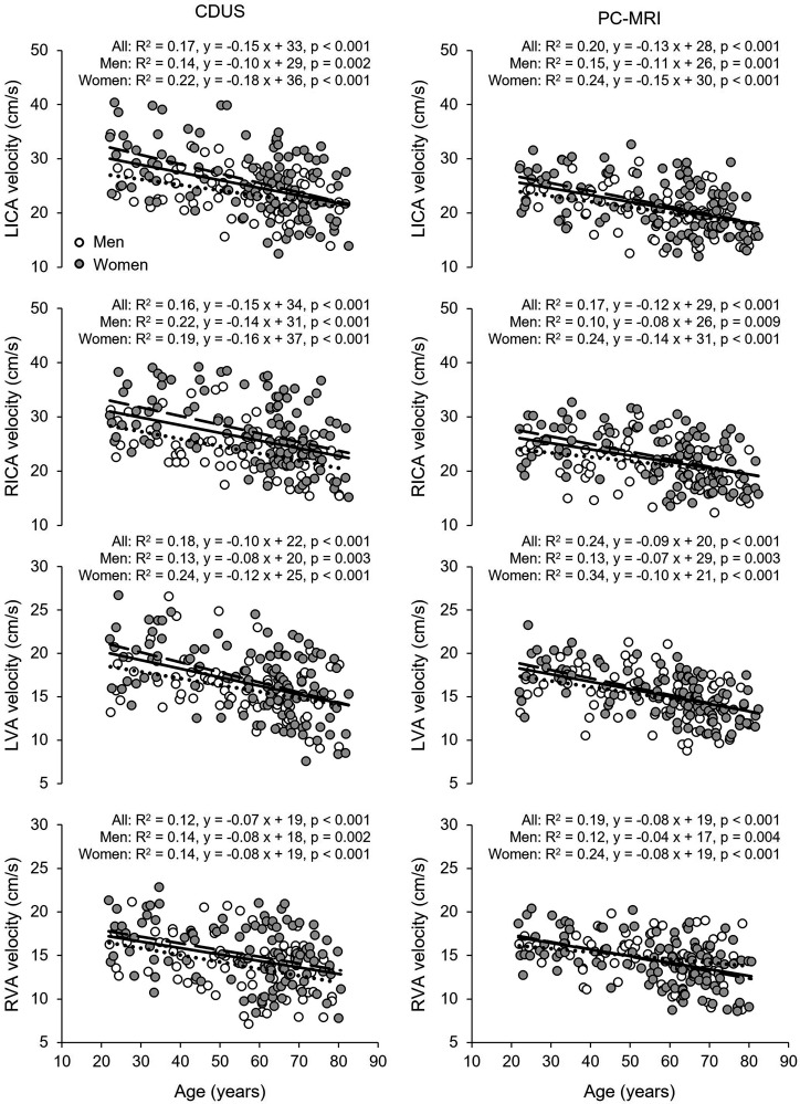
Figure 3.
Association of age with blood flow velocity measured by color-coded duplex ultrasonography (CDUS) (left panels) and phase-contrast MRI (PC-MRI) (right panels) in the left (L) and right (R) internal carotid (ICA) and the vertebral (VA) arteries. Solid line, dotted line, and dashed lines represent the regression equations obtained for all subjects, men, and women, respectively. The rates of decreases in blood flow velocity in the ICA and VA were similar between women and men (p-values of the interaction of age and sex in one-way analysis of covariance: CDUS (left panels) LICA, p = 0.129; RICA, p = 0.622; LVA, p = 0.224; RVA, p = 0.933; PC-MRI (right panels) LICA, p = 0.296; RICA, p = 0.121; LVA, p = 0.222; RVA, p = 0.262).

