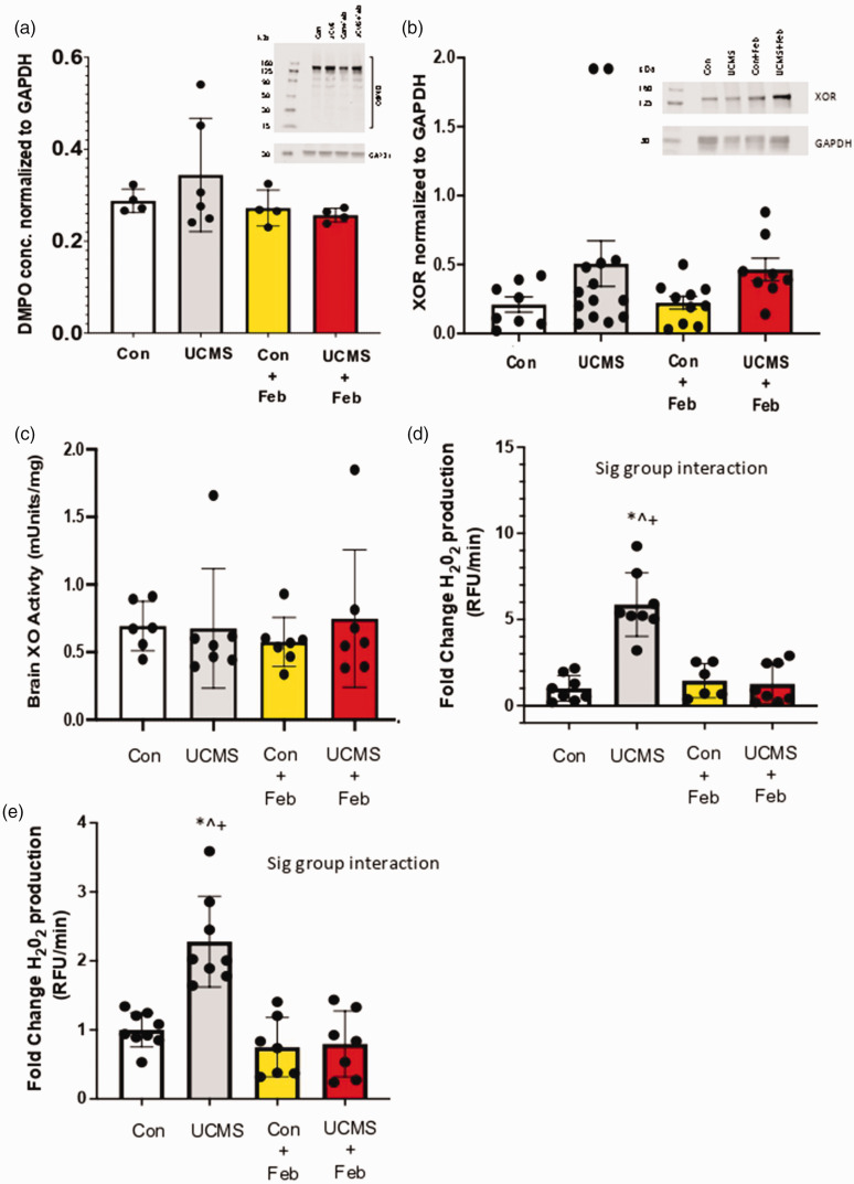
Figure 5.
Oxidative stress in the brain and cerebrovasculature. (a) 5,5-Dimethyl-1-Pyrroline-N-Oxide (DMPO) was used to detect free radical formation on biomolecules in the brain via western blotting; (b) Xanthine oxidase reductase (XOR) concentration was assessed within the liver using western blotting techniques; (c) Brain XO activity was measured with HPLC; Hydrogen peroxide (H2O2) production was measured in whole brain homogenate (d) and isolated brain microvessels (e) musing the CBA assay. *p < 0.05 vs control; ^p < 0.05 vs control + Feb; ‡p < 0.05 vs. UCMS; +p < 0.05 vs UCMS + Feb; Mean ± SD. n = 4–14 mouse tissue samples/group. Two-way ANOVA with a group-by-drug interaction was performed and when a significant interaction (panel D and E) was established a tukey post-hoc test was explored, otherwise, the main variable effect was reported.

