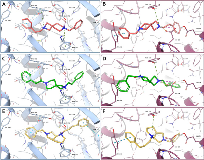
Figure 3.
3D representation of (A, B) compound 4b (salmon carbon sticks), (C, D) compound 5b (green carbon sticks) and (E, F) compound 8f (yellow carbon sticks) in complex with S1R (A, C, E) and S2R (B, D, F), respectively. The S1R and S2R are represented as blue and violet cartoons, respectively. The receptor residues involved in crucial contacts with the compounds are reported as blue and violet carbon sticks. Hydrogen bonds, salt bridges, π–π stacking, and π-cation interactions are represented by yellow, magenta, azure, and green dashed lines, respectively.

