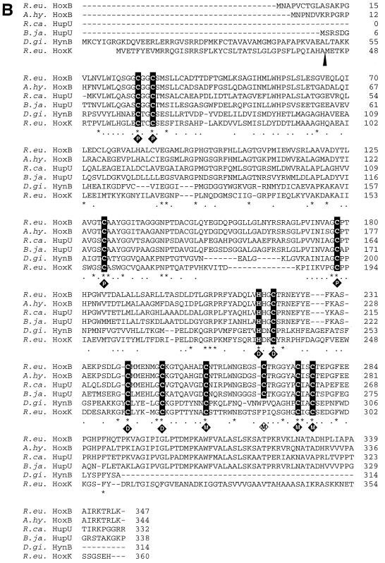
FIG. 2.
Alignment of H2-sensing and standard [NiFe] hydrogenase subunits. Identical and similar amino acids are marked by asterisks and dots, respectively. (A) The five stretches of conservative amino acids identified in [NiFe] hydrogenase large subunits (2) are shown in boldface letters above the sequences (elements 1 to 5). The two cysteine pairs which coordinate nickel and iron at the catalytic center of the D. gigas hydrogenase are highlighted. The cleavage site of C-terminal proteolysis of HynA is marked by an arrowhead. (B) The cysteine and histidine residues which coordinate the proximal (P), medial (M), and distal (D) iron sulfur clusters in the small subunit HynB of D. gigas are highlighted in addition to Cys267 in HoxB of R. eutropha. The cleavage site of the N-terminal leader peptide in HynB is marked by an arrowhead. Abbreviations: R.eu, R. eutropha; A.hy., A. hydrogenophilus; B.ja., B. japonicum; R.ca., R. capsulatus; D.gi., D.gigas.


