Fig. 3.
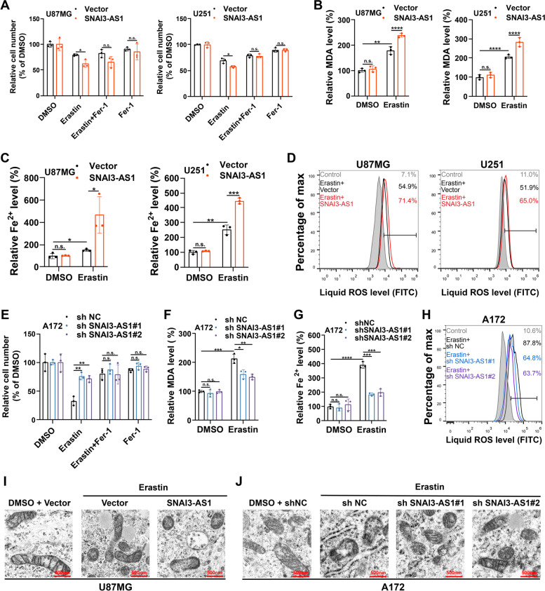
SNAI3-AS1 promotes erastin-induced ferroptosis in vitro. A-D U87MG and U251 cells stably overexpressing SNAI3-AS1 were treated with erastin (10 μM) ± ferrostatin-1 (2 μM) for 48 h, cell viabilities were detected via CCK8 assays (A), intracellular MDA was determined by MDA assays (B), intracellular Fe2+ was measured by iron detection assays (C), lipid ROS accumulation was analyzed by flow cytometry with C11-BODIPY staining (D). E–H A172 cells with stable SNAI3-AS1 knockdown were treated with erastin (10 μM) ± ferrostatin-1 (2 μM) for 48 h, cell viabilities were detected via CCK8 assays (E), intracellular MDA was determined by MDA assays (F), intracellular Fe2+ was measured by iron detection assays (G), lipid ROS accumulation was analyzed by flow cytometry with C11-BODIPY staining (H). Transmission electron microscopy was performed to evaluate the ultrastructural changes of mitochondria in U87MG stably overexpressing SNAI3-AS1 (I) and A172 cells with stable SNAI3-AS1 knockdown (J) after treated with Erastin (10 μM) for 48 h. *P < 0.05, **P < 0.01, ***P < 0.001, ****P < 0.0001, and n.s., not significant

