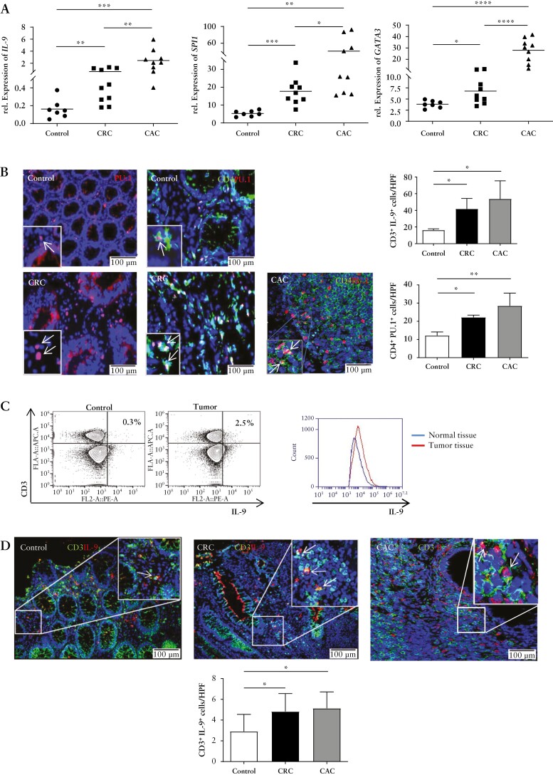
Figure 1.
Elevated expression of PU.1 and IL-9 in patients with colorectal cancer. A] Total mRNA was isolated from patients with colorectal cancer, colitis-associated cancer [CRC, CAC] and healthy controls. Quantitative PCR analysis showed elevated mRNA levels of SPI1, IL9, and GATA3 in tumour tissue. B] Immunofluorescence staining for PU.1 and CD4 from colorectal cancer patients and controls. Indicated cells were counted per high-power field [HPF] [lower panels]. Significant differences are indicated. Data represent mean values ± SEM per high-power field [n = 5–7]. C] FACS analysis of IL-9-expression in T cells in human colonic LPMCs from five tumour and control patients was performed. Representative scatter plots are shown. One representative overlay of IL-9 expression from LPMC T cells of colorectal cancer patients and controls is shown. Gating strategy is represented in the [Supplementary Fig 1]. D] Cryosections from colorectal cancer patients and controls were stained for CD3 and IL-9. Doublepositive cells per HPF and statistical analysis with significant differences [*p < 0.05; **p <0.01] of five patients is shown. CRC, colorectal cancer; CAC, colitis-associated cancer; SEM, standard error of the mean; LPMCs, lamina propria mononuclear cells.

