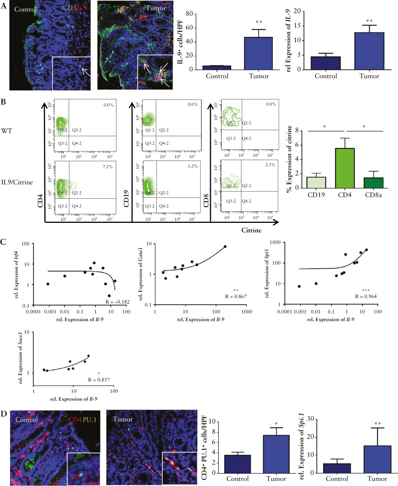
Figure 2.
Th9 cells producing IL-9 are induced in CRC. A] Left panels: immunofluorescence staining for CD3 [green] and IL-9 [red] in the lamina propria of tumour tissue with representative staining, and statistical analysis is shown [n = 3]. Total mRNA from tumour tissue of AOM/DSS-treated wild-type mice and controls was isolated. Right panel: qPCR analysis showed a significant upregulation for IL-9 mRNA levels in experimental CRC [n = 7]. Significant differences are indicated [**p <0.01]. B] Isolated LPMCs from tumour tissue of IL9 citrine reporter mice were stained for CD4, CD19, and CD8. Representative scatter plots are shown from one representative experiment out of two. Statistical analysis with significant differences [n = 3] is shown below [*p <0.05]. C] Relative mRNA expression levels for Il-9 and Gata-3, Spi1, Irf4, and Socs3 were analysed in CRC tissue from wild-type mice. Correlation coefficients are indicated. D] Left panels: immunofluorescence staining for CD4 [red] and PU.1 [green]. Double-positive cells were counted per HPF from six mice [right upper panel]. Right panel: qPCR analysis for the expression levels of Spi1 was performed on tumour tissue and control tissue [n = 10] from wild-type mice. CRC, colorectal cancer, LPMCs, lamina propria mononuclear cells; HPF, high-power field.

