Figure 7.
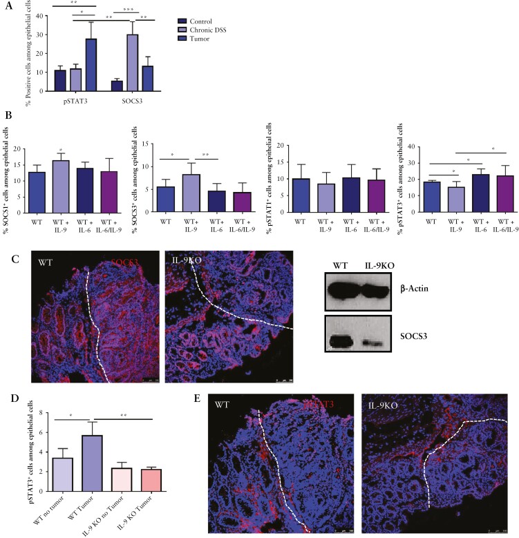
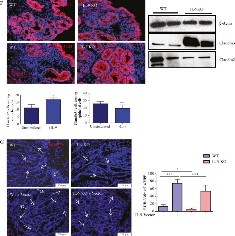
IL-9 effects on epithelial cells of colitis-associated neoplasias. A] Assessment of SOCS3 and pSTAT3 expression during CAC. Isolated epithelial cells from WT mice were investigated by FACS during chronic DSS inflammation and after tumour formation and compared with healthy controls. Results from four mice per condition and significant differences are indicated [*p <0.05; **p <0.01; ***p <0.001]. B] FACS analysis of organoids incubated with either 100 ng IL-9, 100 ng IL-6, or the combination of both cytokines. Organoids were stained for the epithelial marker EpCAM and the intracellular markers SOCS1, SOCS3, pSTAT1, or pSTAT3. Statistical analysis of six different experiment is shown [*p <0.05; **p <0.01]. C] Left panels: cryosections from AOM/DSS-treated wild-type and IL-9 knockout mice were stained for the expression of SOCS3. Nuclei were stained with DAPI. Representative stainings of three mice per group are shown. Right panel: isolated epithelial cells from tumour tissue of wild-type and IL-9 KO mice were analysed by western blot for SOCS3 expression with β-Actin as loading control. D] Isolated epithelial cells of tumour tissue and normal tissue from wild-type and IL-9-deficient mice were analysed by FACS for the expression of pSTAT3. Statistical analysis with significant differences [*p <0.05; **p <0.01] is shown [four mice per group]. E] Cryosections from wild-type and IL-9 knockout mice were stained with an anti-pSTAT3 antibody and cell nuclei with DAPI. pSTAT3+ cells were detected mostly in the tumour tissue of wild-type mice. Representative stainings are shown [three mice per group]. F] Expression of claudin tight junctions was analysed on cryosections of tumour tissue from wild-type and IL-9 knockout mice. Representative pictures are shown for claudin-2 [Cld2] and claudin-3 [Cld3] [left panels]. Epithelial cells from tumour tissue of wild-type and IL-9 deficient mice were analysed by western blots for the protein expression of claudins with β-Actin as loading control [right panel]. G] FISH analysis for the assessment of bacterial translocation in wild-type and IL-9 knockout mice. Cryosections were stained with the EUB-338 probe to detect bacteria [arrows]. Representative stainings are shown from one mouse out of four mice per group. Statistical analysis of EUB-338+ cells per HPF is shown [*p <0.05; ***p <0.001]. CAC, colitis-associated cancer; AOM, azoxymethane; DSS, dextran sodium sulphate; WT, wild-type; HPF, high-power field.


